Differential activation in somatosensory cortex for different discrimination tasks
- PMID: 10627620
- PMCID: PMC6774136
- DOI: 10.1523/JNEUROSCI.20-01-00446.2000
Differential activation in somatosensory cortex for different discrimination tasks
Abstract
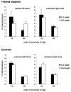
Maps of the body surface in somatosensory cortex have been shown to be highly plastic, altering their configuration in response to changes in use of body parts. The current study investigated alterations in the functional organization of the human somatosensory cortex resulting from massed practice. Over a period of 4 weeks, subjects were given synchronous tactile stimulation of thumb (D1) and little finger (D5) for 1 hr/d. They had to identify the orientation of the stimuli. Neuroelectric source localization based on high-resolution EEG revealed that, when subjects received passive tactile stimulation of D1 or D5, the representations of the fingers in primary somatosensory cortex were closer together after training than before. There was also an apparently correlative tendency to anomalously mislocalize near-threshold tactile stimuli equally to the distant finger costimulated during training rather than preferentially to the finger nearest to the finger stimulated in a post-training test. However, when the stimulus discrimination had to be made, neuroelectric source imaging revealed that the digital representations of D1 and D5 were further apart after training than before. Thus, the same series of prolonged repetitive stimulations produced two different opposite effects on the spatial relationship of the cortical representations of the digits, suggesting that differential activation in the same region of somatosensory cortex is specific to different tasks.
Figures


References
-
- Allard T, Clark SA, Jenkins WM, Merzenich MM. Reorganization of somatosensory area 3b representations in adult owl monkeys after digital syndactyly. J Neurophysiol. 1991;66:1048–1058. - PubMed
-
- Baumgartner C, Doppelbauer A, Sutherling WW, Lindinger G, Levesque MF, Aull S, Zeitlhofer J, Deecke L. Somatotopy of human somatosensory cortex as studied in scalp EEG. Electroencephalogr Clin Neurophysiol. 1993;88:271–279. - PubMed
-
- Braun C, Kaiser S, Kincses WE, Elbert T. Confidence interval of single dipole locations based on EEG data. Brain Topogr. 1997;10:31–39. - PubMed
-
- Buonomano DV, Merzenich MM. Cortical plasticity: from synapses to maps. Annu Rev Neurosci. 1998;21:149–186. - PubMed
-
- Clark SA, Allard T, Jenkins WM, Merzenich MM. Receptive fields in the body surface map in adult cortex defined by temporally correlated inputs. Nature. 1988;332:444–445. - PubMed
Publication types
MeSH terms
LinkOut - more resources
Full Text Sources
