. 2000 Feb;20(3):755-9.
doi: 10.1128/MCB.20.3.755-759.2000.

Normal development, wound healing, and adenovirus susceptibility in beta5-deficient mice

Affiliations

Normal development, wound healing, and adenovirus susceptibility in beta5-deficient mice

X Huang et al. Mol Cell Biol. 2000 Feb.

Abstract

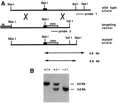
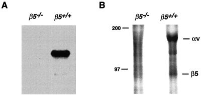
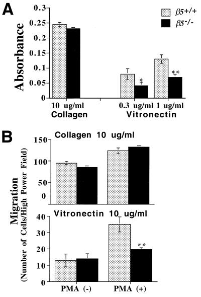
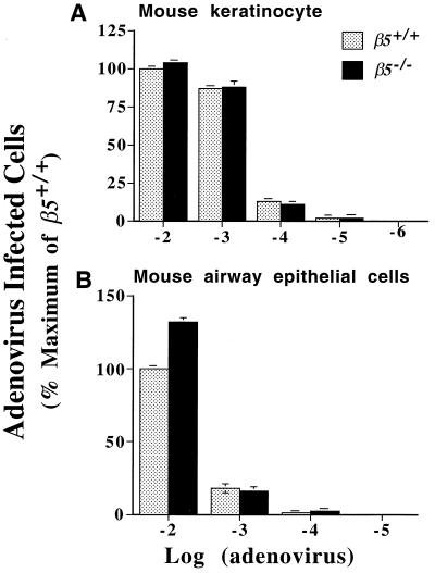
Integrins have been shown to play important roles in embryonic development, wound healing, metastasis, and other biological processes. alphavbeta5 is a receptor for RGD-containing extracellular matrix proteins that has been suggested to be important in cutaneous wound healing and adenovirus infection. To examine the in vivo function of this receptor, we have generated mice lacking beta5 expression, using homologous recombination in embryonic stem cells. Mice homozygous for a null mutation of the beta5 subunit gene develop, grow, and reproduce normally. Keratinocytes harvested from beta5(-/-) mice demonstrate impaired migration on and adhesion to the alphavbeta5 ligand, vitronectin. However, the rate of healing of cutaneous wounds is not different in beta5(-/-) and beta5(+/+) mice. Furthermore, keratinocytes and airway epithelial cells obtained from null mice show adenovirus infection efficiency equal to that from wild-type mice. These data suggest that alphavbeta5 is not essential for normal development, reproduction, adenovirus infection, or the healing of cutaneous wounds.


Figures

FIG. 1
FIG. 1
(A) Homologous recombination of the β5 gene in mouse ES cells. Top panel, 8.2 kb of normal genomic structure of the mouse β5 gene. One exon is shown as a solid box. Middle panel, the targeting plasmid in pBluescript is linearized at a unique SacII site. Neomycin resistance (neo) and herpes simplex virus thymidine kinase (TK) genes are shown as shaded boxes. Bottom panel, structure of the β5 gene after a correct targeting event. (B) Southern blot analysis of ES clones. SpeI-digested genomic DNA was blotted with the external probe (probe 1). The external probe gives a 4.6-kb band for the wild-type allele and a 3.8-kb band for the mutant allele since the neomycin resistance gene is shorter than the fragment replaced.
FIG. 2
FIG. 2
(A) Northern analysis of mRNA from β5+/+ and β5−/− mice. Total RNA was extracted from cultured keratinocytes and transcribed to cDNA. The specific cDNA probe of mouse β5 was used to detect message from wild-type but not deficient cells. (B) Immunoprecipitation of αvβ5 from wild-type but not β5−/− murine keratinocytes. [35S]methionine-labeled keratinocyte lysate was immunoprecipitated with anti-β5 antibody raised against the human β5 cytoplasmic domain. Immunoprecipitated proteins were analyzed by SDS-PAGE under nonreducing conditions. The positions of molecular mass marker (in kilodaltons) are shown to the left.
FIG. 3
FIG. 3
(A) Cell adhesion of murine keratinocytes. Confluent keratinocytes from β5−/− and wild-type mice were harvested with trypsin and plated on 96-well plates coated with collagen or vitronectin. Cells were allowed to attach to the wells for an hour and were then fixed and stained. Adhesion was expressed as absorbance at 595 nm. Data calculated from triplicate wells were expressed as the mean (± standard error of the mean). Significantly less than wild type: *, P < 0.05; ∗∗, P < 0.01. (B) Cell migration of murine keratinocytes. Cells were harvested as described above and plated on transwell membranes coated with collagen (10 μg/ml) or vitronectin (10 μg/ml) in the presence or absence of PMA. Cells that had migrated onto the bottom side of the membrane were stained and counted by inverted microscopy at 40×. The data were expressed as the mean (± standard error of the mean). ∗∗, Significantly less than wild type, P < 0.01.
FIG. 4
FIG. 4
Keratinocytes (A) and airway epithelial cells (B) from wild-type and β5−/− mice were infected with a range of concentrations of adenovirus H5.010CMVlacZ; 40 h after infection, cells were treated with X-Gal and blue-stained cells were counted. The results are expressed as a percentage of the number of positive wild-type cells after incubation with the highest concentration of adenovirus used. Results are the mean (± standard error of the mean) of three experiments.

References

    1. Adams J C, Watt F M. Expression of beta 1, beta 3, beta 4, and beta 5 integrins by human epidermal keratinocytes and non-differentiating keratinocytes. J Cell Biol. 1991;115:829–841. - PMC - PubMed
    1. Bader B L, Rayburn H, Crowley D, Hynes R O. Extensive vasculogenesis, angiogenesis, and organogenesis precede lethality in mice lacking all alpha v integrins. Cell. 1998;95:507–519. - PubMed
    1. Breuss J M, Gillett N, Lu L, Sheppard D, Pytela R. Restricted distribution of integrin beta 6 mRNA in primate epithelial tissues. J Histochem Cytochem. 1993;41:1521–1527. - PubMed
    1. Busk M, Pytela R, Sheppard D. Characterization of the integrin αvβ6 as a fibronectin-binding protein. J Biol Chem. 1992;267:5790–5796. - PubMed
    1. Capecchi M R. Altering the genome by homologous recombination. Science. 1989;244:1288–1292. - PubMed

Publication types

MeSH terms

LinkOut - more resources