. 2000 Feb;20(3):816-24.
doi: 10.1128/MCB.20.3.816-824.2000.

Biochemical and genetic conservation of fission yeast Dsk1 and human SR protein-specific kinase 1

Affiliations

Biochemical and genetic conservation of fission yeast Dsk1 and human SR protein-specific kinase 1

Z Tang et al. Mol Cell Biol. 2000 Feb.

Abstract

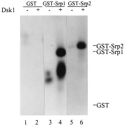
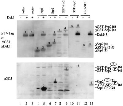
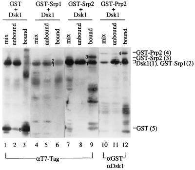
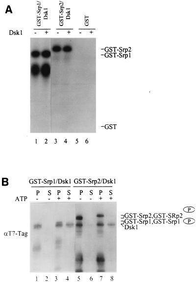
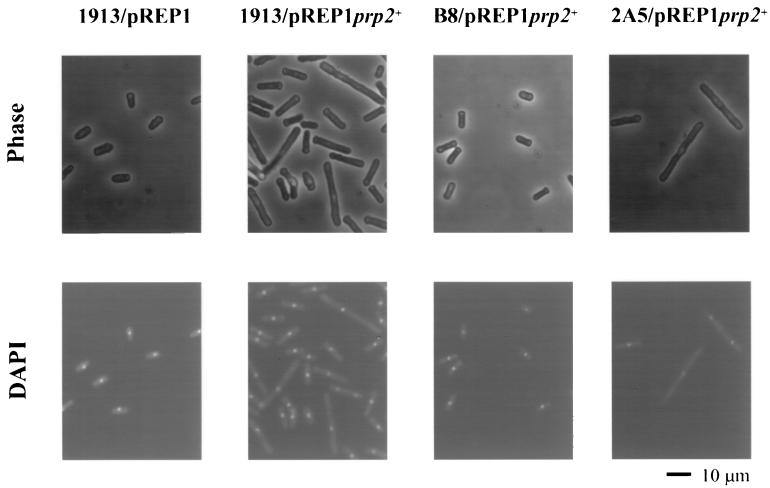
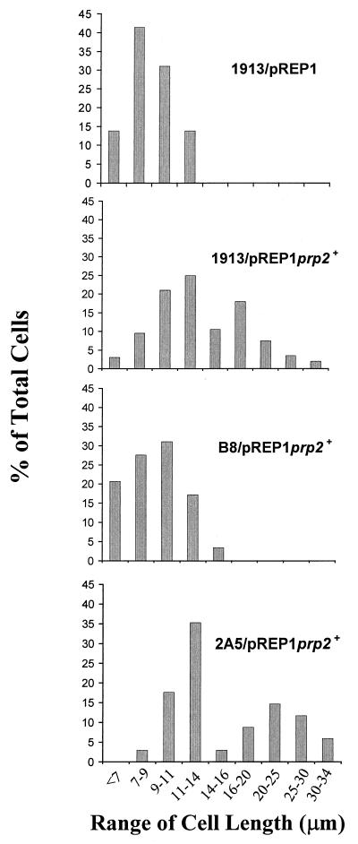
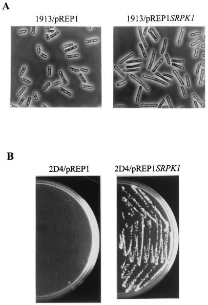
Arginine/serine-rich (RS) domain-containing proteins and their phosphorylation by specific protein kinases constitute control circuits to regulate pre-mRNA splicing and coordinate splicing with transcription in mammalian cells. We present here the finding that similar SR networks exist in Schizosaccharomyces pombe. We previously showed that Dsk1 protein, originally described as a mitotic regulator, displays high activity in phosphorylating S. pombe Prp2 protein (spU2AF59), a homologue of human U2AF65. We now demonstrate that Dsk1 also phosphorylates two recently identified fission yeast proteins with RS repeats, Srp1 and Srp2, in vitro. The phosphorylated proteins bear the same phosphoepitope found in mammalian SR proteins. Consistent with its substrate specificity, Dsk1 forms kinase-competent complexes with those proteins. Furthermore, dsk1(+) gene determines the phenotype of prp2(+) overexpression, providing in vivo evidence that Prp2 is a target for Dsk1. The dsk1-null mutant strain became severely sick with the additional deletion of a related kinase gene. Significantly, human SR protein-specific kinase 1 (SRPK1) complements the growth defect of the double-deletion mutant. In conjunction with the resemblance of dsk1(+) and SRPK1 in sequence homology, biochemical properties, and overexpression phenotypes, the complementation result indicates that SRPK1 is a functional homologue of Dsk1. Collectively, our studies illustrate the conserved SR networks in S. pombe consisting of RS domain-containing proteins and SR protein-specific kinases and thus establish the importance of the networks in eucaryotic organisms.


Figures

FIG. 1
FIG. 1
Dsk1 phosphorylates fission yeast Srp1 and Srp2 proteins in vitro. GST fusion proteins were isolated from bacterial lysates by binding to glutathione beads. After a washing, the bound GST (lanes 1 and 2), GST-Srp1 (lanes 3 and 4), and GST-Srp2 (lanes 5 and 6) were individually incubated with purified Dsk1 (lanes 2, 4 and 6) in the presence of [γ-32P]ATP at 23°C for 30 min. Samples were resolved on an SDS–10% polyacrylamide gel and visualized with X-ray film. The expected positions of GST, GST-Srp1, and GST-Srp2 proteins on the gel are indicated on the right. Truncated forms of GST-Srp1 protein were observed as a lower-molecular-size band (lane 4).
FIG. 2
FIG. 2
Dsk1-mediated phosphorylation of Srp1, Srp2, and Prp2 proteins generates a phosphoepitope specifically recognized by MAb 3C5. Purified GST-Prp2 (lanes 10 and 11), purified GST-SF2/ASF (lanes 12 and 13), or a bacterial lysate containing individual recombinant proteins (lanes 4 to 9) as indicated at the top of each lane was incubated with (lanes 5, 7, 9, 11, and 13) or without (lanes, 4, 6, 8, 10, and 12) purified Dsk1 protein in the presence of an ATP regenerating system for 30 min at 23°C. Buffer (lanes 1 and 2) and lysate from bacteria with the pET28a vector alone (lane 3) were used as negative controls. The samples were then processed for immunoblotting with anti-T7-Tag MAb (top panel, lanes 1 to 9) or anti-GST and anti-Dsk1 polyclonal antibodies in successive order (top panel, lanes 10 to 13) or MAb 3C5 monoclonal antibody (bottom panel). Alkaline phosphatase-conjugated goat anti-mouse immunoglobulin G (IgG) and goat anti-rabbit (top panel) or goat anti-mouse (bottom panel) IgM antibodies were used as secondary antibodies. The identity of the proteins is marked above each band with numbers 1 to 6 representing Dsk1, Srp1, Srp2, GST-Srp2, GST-Prp2, and GST-SF2, respectively, as indicated on the right side of the figure. The same amount of Srp1 and Srp2 was used, while GST-Srp2 at 1/4 of the amount and GST-Prp2 and GST-SF2/ASF at <1/10 of the amount were added to the indicated samples.
FIG. 3
FIG. 3
Srp1, Srp2, and Prp2 proteins individually form a complex with Dsk1 in vitro. A bacterial lysate containing Dsk1 protein was incubated with a lysate containing GST (lanes 1 to 3) or GST fusion (lanes 4 to 12) proteins as indicated at the top of each lane to allow complex formation at 23°C for 30 min. Glutathione beads were then added to pulldown bound proteins at 4°C as described in Materials and Methods. Portions of mixed lysates, unbound fractions, and bound fractions from each sample were analyzed by SDS-polyacrylamide gel electrophoresis. Some samples were processed for immunoblotting by using anti-T7-Tag MAb (lanes 1 to 9), which detects GST, GST-Srp1, GST-Srp2, and Dsk1. Other samples were processed for immunoblotting first with anti-GST and subsequently with anti-Dsk1 polyclonal antibodies (lanes 10 to 12). Dsk1 protein was pulled down by each of the four RS domain-containing proteins (lanes 6, 9, and 12) but not by GST protein (lane 3). Numbers 1 to 5 on the left of the protein bands in the bound fraction of each sample represent Dsk1, GST-Srp1, GST-Srp2, GST-Prp2, and GST, respectively, as indicated on the right side of the figure.
FIG. 4
FIG. 4
Dsk1 is dissociated from the complex after phosphorylation of Srp1 or Srp2 protein. (A) The bound Dsk1 phosphorylates Srp1 and Srp2 in the complex in the presence of ATP. The pulldown complexes GST-Srp1/Dsk1 (lanes 1 and 2) and GST-Srp2/Dsk1 (lanes 3 and 4), as described in Fig. 3, were incubated with (lanes 2 and 4) or without (lanes 1 and 3) purified Dsk1 protein in the presence of [γ-32P]ATP. GST protein was also used in place of the GST fusion proteins as a negative control (lanes 5 and 6). Samples were resolved on an SDS–10% polyacrylamide gel and visualized by autoradiography. The bound Dsk1 phosphorylated Srp1 and Srp2 in the complex (lanes 1 and 3). (B) After the kinase reaction, Dsk1 is released from the Srp1/Dsk1 and Srp2/Dsk1 complexes. GST-Srp1/Dsk1 and GST-Srp2/Dsk1 protein complexes were incubated individually with (lanes 3, 4, 7, and 8) or without (lanes 1, 2, 5, and 6) an ATP regenerating system for 30 min at 23°C. Following the kinase reaction, protein-bound beads were pelleted by centrifugation. The supernatant (S) and bead (P) portions of each sample were resolved on an SDS–10% polyacrylamide gel and subsequently processed for immunoblotting with anti-T7-Tag MAb. Dsk1 was released from the complex to the supernatant in the presence of ATP (lanes 4 and 8), but it is not dissociated from the complex in the absence of ATP (lanes 2 and 6). Note the phosphorylated Srp proteins (indicated with a circled P) have slower mobility than that of their nonphosphorylated forms.
FIG. 5
FIG. 5
The cell elongation phenotype resulting from Prp2 overproduction is dependent on the dsk1+ gene. Strains 1913 (wild type), B8 (Δdsk1), and 2A5 (Δkic1) were transformed with pREP1prp2+. Strain 1913 containing pREP1 vector was also generated as a negative control. Cells were first grown at 32°C to midlogarithmic phase in minimal medium (EMM2) with thiamine, and Prp2 overproduction was then induced for 21 h in the absence of thiamine. Cells were fixed by heating them on slides, and they were then stained with DAPI. Cell images obtained by phase-contrast (top panel) and fluorescence (bottom panel) microscopy were indicated. Magnification is ×400 in all panels. The elongated cells were observed in strain 1913 (wild type, second column) and 2A5 (Δkic1, fourth column) but not in strain B8 (Δdsk1, third column). The scale bar represents 10 μm.
FIG. 6
FIG. 6
Size distribution of cell population in strains with prp2+ gene overexpressed. The cell length of the four samples in Fig. 5 was measured. The cell populations with a size range as indicated for 1913/pREP1, 1913/pREP1prp2+, B8/pREP1prp2+, and 2A5/pREP1prp2+ are displayed as histograms. A population of cells longer than 16 μm was observed in 1913/pREP1prp2+ and 2A5/pREP1prp2+. The distribution pattern of 2A5/pREP1prp2+ is similar to that 1913/pREP1prp2+, while the pattern of B8/pREP1prp2+ resembles that of the negative control, 1913/pREP1.
FIG. 7
FIG. 7
Human SRPK1 is a functional homologue of fission yeast Dsk1. (A) Overexpression of human SRPK1 gene in S. pombe results in elongated cells similar to the cells with dsk1+ overexpression. Strain 1913 (wild type) was transformed with either pREP1 or pREP1SRPK1. Exponentially grown cells were induced for 16 to 18 h in the absence of thiamine and fixed for microscopy. Left panel and right panel show at a magnification of ×400 the phase-contrast micrographs of cells harboring pREP1 and pREP1SRPK1, respectively. Fission yeast cells with human SRPK1 gene overexpressed were elongated (right panel) compared to those carrying pREP1 vector alone (left panel) under the same condition. (B) Expression of human SRPK1 gene complements the growth defect of Δdsk1Δkic1 double-deletion strain (2D4). Strain 2D4 (Δdsk1Δkic1) was transformed with either pREP1 or pREP1SRPK1. The transformants were subsequently analyzed on minimal medium plates in the presence of thiamine and incubated for 4 days at 33°C. Cells carrying pREP1SRPK1 formed healthy colonies (right panel), whereas cells harboring pREP1 hardly grew (left panel) under the same condition.

Similar articles

Cited by

References

    1. Alfa C, Fantes P, Hyams J, McLeod M, Warbrick E. Experiments with fission yeast: a laboratory course manual. Cold Spring Harbor, N.Y: Cold Spring Harbor Laboratory Press; 1993.
    1. Bridge E, Xia D X, Carmo-Fonseca M, Cardinali B, Lamond A I, Pettersson U. Dynamic organization of splicing factors in adenovirus-infected cells. J Virol. 1995;69:281–290. - PMC - PubMed
    1. Cao W, Jamison S F, Garcia-Blanco M A. Both phosphorylation and dephosphorylation of ASF/SF2 are required for pre-mRNA splicing in vitro. RNA. 1997;3:1456–1467. - PMC - PubMed
    1. Colwill K, Feng L L, Yeakley J M, Gish G D, Caceres J F, Pawson T, Fu X D. SRPK1 and Clk/Sty protein kinases show distinct substrate specificities for serine/arginine-rich splicing factors. J Biol Chem. 1996;271:24569–24575. - PubMed
    1. Colwill K, Pawson T, Andrews B, Prasad J, Manley J L, Bell J C, Duncan P I. The Clk/Sty protein kinase phosphorylates SR splicing factors and regulates their intranuclear distribution. EMBO J. 1996;15:265–275. - PMC - PubMed

Publication types

MeSH terms