. 2000 Feb;20(3):851-67.
doi: 10.1128/MCB.20.3.851-867.2000.

The Cbl proto-oncogene product negatively regulates the Src-family tyrosine kinase Fyn by enhancing its degradation

Affiliations

The Cbl proto-oncogene product negatively regulates the Src-family tyrosine kinase Fyn by enhancing its degradation

C E Andoniou et al. Mol Cell Biol. 2000 Feb.

Abstract

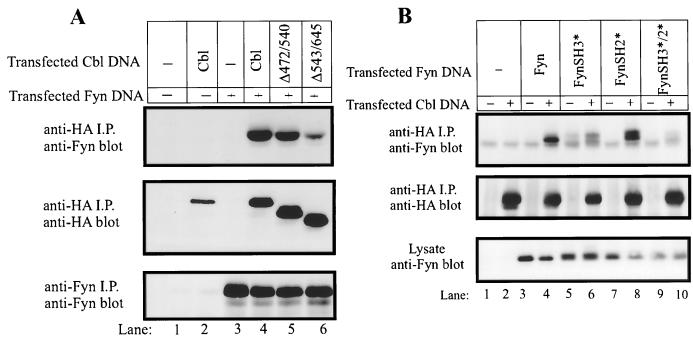
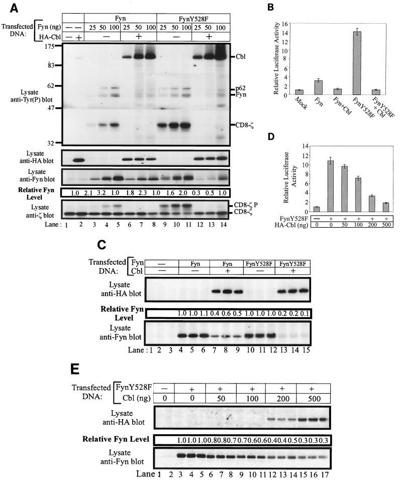
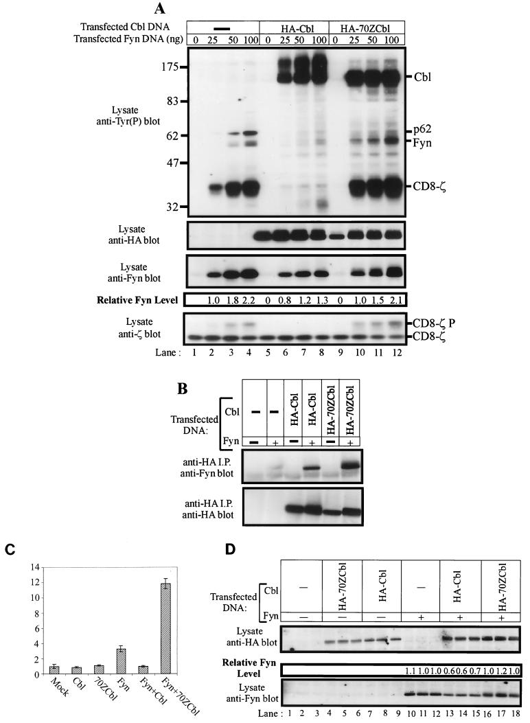
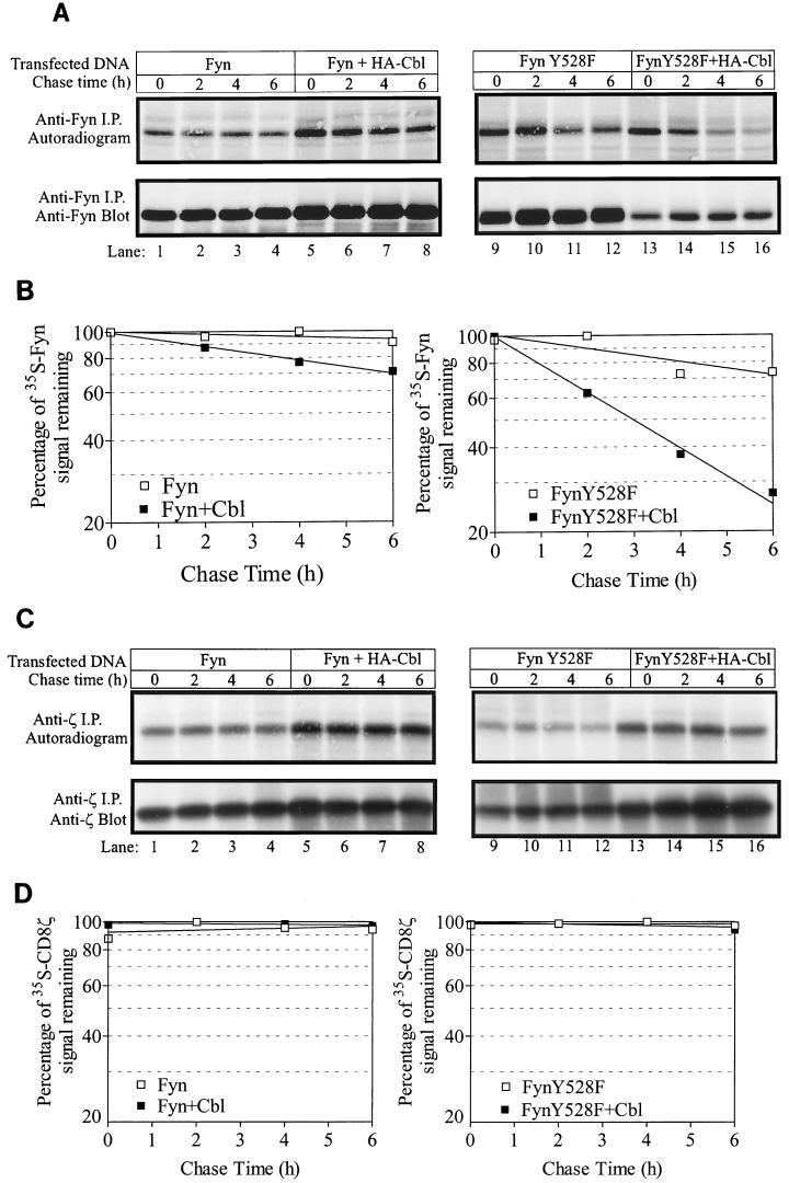
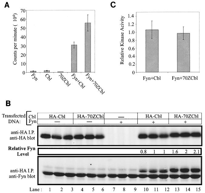
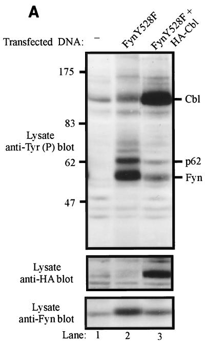
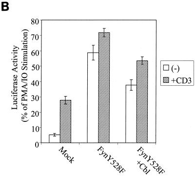
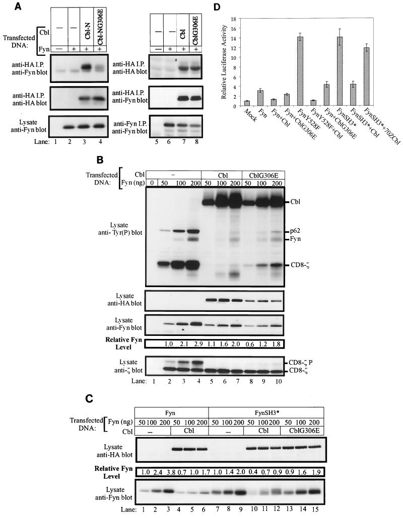
Fyn is a prototype Src-family tyrosine kinase that plays specific roles in neural development, keratinocyte differentiation, and lymphocyte activation, as well as roles redundant with other Src-family kinases. Similar to other Src-family kinases, efficient regulation of Fyn is achieved through intramolecular binding of its SH3 and SH2 domains to conserved regulatory regions. We have investigated the possibility that the tyrosine kinase regulatory protein Cbl provides a complementary mechanism of Fyn regulation. We show that Cbl overexpression in 293T embryonic kidney and Jurkat T-lymphocyte cells led to a dramatic reduction in the active pool of Fyn; this was seen as a reduction in Fyn autophosphorylation, reduced phosphorylation of in vivo substrates, and inhibition of transcription from a Src-family kinase response element linked to a luciferase reporter. Importantly, a Fyn mutant (FynY528F) relieved of intramolecular repression was still negatively regulated by Cbl. The Cbl-dependent negative regulation of Fyn did not appear to be mediated by inhibition of Fyn kinase activity but was correlated with enhanced protein turnover. Consistent with such a mechanism, elevated levels of Fyn protein were observed in cell lines derived from Cbl(-/-) mice compared to those in wild-type controls. The effects of Cbl on Fyn were not observed when the 70ZCbl mutant protein was analyzed. Taken together, these observations implicate Cbl as a component in the negative regulation of Fyn and potentially other Src-family kinases, especially following kinase activation. These results also suggest that protein degradation may be a general mechanism for Cbl-mediated negative regulation of activated tyrosine kinases.


Figures

FIG. 1
FIG. 1
Role of the Cbl proline-rich region and Fyn SH3 domain in Cbl-Fyn association in vivo. (A) 293T cells in 100-mm tissue culture dishes were transfected with the AprM8 expression vector encoding Fyn (0.5 μg), with or without pSRαneo expression vectors (0.5 μg) encoding HA-tagged Cbl constructs. Cell lysates were prepared 48 h after transfection, and 1-mg aliquots of lysate were subjected to immunoprecipitation (i.p.) with anti-HA or anti-Fyn antibodies as indicated. Immune complexes were resolved by SDS-PAGE, transferred to a PVDF membrane, and immunoblotted with anti-HA (middle panel) or anti-Fyn (top and bottom panels) antibodies. (B) 293T cells were transfected with the indicated pAlterMAX-Fyn constructs (0.1 μg), with or without pAlterMAX-HA-Cbl (1 μg). Anti-HA immunoprecipitations (i.p.) were carried out with 200 μg of cell lysate protein. Immune complexes or 15 μg of whole-cell lysate proteins were resolved by SDS-PAGE, transferred to a PVDF membrane, and blotted with the indicated antibodies.
FIG. 2
FIG. 2
Overexpression of Cbl decreases the phosphorylation of in vivo Fyn substrates, reduces the steady-state pools of Fyn proteins, and inhibits the Fyn- or FynY528F-dependent transactivation of an SRE-luciferase reporter. (A) 293T cells were transfected with the indicated amounts of pAlterMAX-Fyn or FynY528F and 0.5 μg of pSRαneo-CD8-ζ, with or without the pAlterMAX-HA-Cbl expression construct (1 μg). Cell lysates were prepared 48 h posttransfection, and 15-μg aliquots of cell lysate were resolved by SDS-PAGE, transferred to a PVDF membrane, and immunoblotted with the indicated antibodies. Fyn protein levels were determined as described in Materials and Methods. This experiment was performed three times, with similar results in each case. (B) 293T cells were transfected with the SRE-luciferase reporter construct (5 μg), together with pSRαneo-CD8-ζ (0.5 μg), pAlterMAX-Fyn or FynY528F (0.1 μg), and pAlterMAX-HA-Cbl (1 μg) expression plasmids as indicated. At 48 h posttransfection, the cells were lysed and equal amounts of protein were used to assay luciferase activity. Results are shown as the mean and 1 standard deviation (SD) based on five replicate transfections, and are expressed relative to the luciferase activity of the mock transfectant, which was arbitrarily set at 1. (C) Fyn and Cbl protein expression from panel B was determined by separating 7.5-μg aliquots of three replicates by SDS-PAGE, transferring them to a PVDF membrane, and immunoblotting with the appropriate antibodies. Relative Fyn protein levels are shown above the Fyn immunoblot. (D) 293T cells were transfected with pAlterMAX-FynY528F (0.1 μg), pSRαneo-CD8-ζ (0.5 μg), and the SRE-luciferase (5 μg) constructs, along with the indicated amounts of pAlterMAX-HA-Cbl. At 48 h posttransfection, the cells were lysed and SRE-luciferase activity was measured as in panel B. Results are shown as the mean and 1 SD based on five replicate transfections and are expressed relative to the luciferase activity of the mock transfectant, which was arbitrarily set at 1. (E) Aliquots (15 μg) of the cell lysates used in panel D were resolved by SDS-PAGE, transferred to a PVDF membrane, and immunoblotted with either anti-HA (top panel) or anti-Fyn (bottom panel) antibodies. The relative Fyn protein levels are shown above the Fyn immunoblot. Tyr(P), phosphotyrosine.
FIG. 3
FIG. 3
70ZCbl enhances Fyn autophosphorylation, phosphorylation of in vivo substrates, and SRE-luciferase transactivation. (A) The indicated amounts of pAlterMax-Fyn and 0.5 μg of pSRαneo-CD8-ζ were transfected into 293T cells, either alone or in the presence of 1 μg of pAlterMax-HA-Cbl or pAlterMAX-HA- 70ZCbl. Aliquots (10 μg) of cell lysates were separated by SDS-PAGE, transferred to a PVDF membrane, and immunoblotted with the indicated antibodies. Relative Fyn protein levels were determined as described in Materials and Methods. (B) Lysates (250 μg) from panel A (lanes 1, 4, 5, 8, 9, and 12) were subjected to anti-HA immunoprecipitation (i.p.). Bound proteins were separated by SDS-PAGE, transferred to a PVDF membrane, and blotted with either anti-HA or anti-Fyn antibodies. (C) 293T cells were transfected with the SRE-luciferase reporter construct (5 μg), pSRαneo-CD8-ζ (0.5 μg), and the indicated combinations of pAlterMAX-Fyn (0.1 μg), pAlterMAX-HA-Cbl, or pAlterMAX-HA-70ZCbl (1 μg) expression plasmids. At 48 h posttransfection, the cells were lysed and equal amounts of protein were used to assay luciferase activity. Luciferase activity was determined, and the results were expressed relative to the luciferase activity of the mock transfectant, which was arbitrarily set at 1. Results represent the mean and 1 SD of five replicate transfections. (D) Aliquots (10 μg) of the cell lysates used in panel C were resolved by SDS-PAGE, transferred to a PVDF membrane, and immunoblotted with either anti-HA (top panel) or anti-Fyn (bottom panel) antibodies. Relative Fyn protein levels are shown above the anti-Fyn immunoblot. Tyr(P), phosphotyrosine.
FIG. 4
FIG. 4
Cbl overexpression enhances the turnover of Fyn and FynY528F protein as determined by metabolic pulse-chase analysis but has no effect on the half-life of CD8-ζ. 293T cells in 100-mm tissue culture dishes were transfected with pSRαneo-CD8-ζ (0.5 μg), pAlterMAX-Fyn (0.1 pg), or FynY528F (0.1 μg) with or without the pAlterMAX-HA-Cbl expression construct (1 μg). At 48 h posttransfection, the cells were methionine starved for 1 h and then were pulse-labeled with [35S]methionine for 45 min. The pulse was chased with culture medium supplemented with unlabeled methionine for the indicated times, and cell lysates were prepared. (A) Anti-Fyn immunoprecipitates (i.p.) of cell lysates were resolved by SDS-PAGE and transferred to a PVDF membrane. Labeled Fyn protein was visualized by exposing the membrane to X-ray film for 48 h (top panel). The membrane was subsequently immunoblotted with an anti-Fyn antibody (bottom panel). (B) The signals in panel A were quantified as described in Materials and Methods, and the values at various chase times are expressed as a percentage relative to the highest 35S-Fyn signal. The line of best fit was calculated with the Prism program. A representative experiment is shown. This experiment was performed three times, with similar results in each case. (C) CD8-ζ protein was immunoprecipitated (i.p.) from lysates prepared by the same method as in panel A, and the labeled protein was visualized by autoradiography following transfer to PVDF (top panel, 16-h exposure). The membrane was then immunoblotted with a ζ-specific antibody (bottom panel). (D) Signals in panel C were quantified and expressed as for panel B.
FIG. 5
FIG. 5
Cbl-associated Fyn kinase activity does not differ from 70ZCbl-associated Fyn activity. (A and B) 293T cells were transfected with either 1 μg of pAlterMAX-HA-Cbl or pAlterMAX-HA-70ZCbl alone or in combination with 0.05 μg of pAlterMAX Fyn. The cells were lysed 48 h after transfection, and Cbl proteins were immunoprecipitated from 900 μg of lysate with the 12CA5 anti-HA antibody. Following washing, the immunoprecipitates (i.p.) were divided equally and used for either an in vitro kinase assay (A) or immunoblot analysis (B). (A) Kinase activity associated with Cbl or 70ZCbl was determined by incubating the bound proteins in kinase buffer containing a synthetic substrate (Raytide) and [γ32P]ATP. 32P incorporation into the Raytide substrate was quantified with a Packard 1600TR liquid scintillation counter. Results are expressed as the mean and 1 SD of three replicates. (B) Fyn associated with Cbl or 70ZCbl was visualized by immunoblotting and quantified as described in Materials and Methods. Data from triplicate samples are shown for each transfection condition. (C) Fyn kinase activities in panel A were normalized for the Fyn protein levels observed in panel B. This experiment was conducted twice, with similar results observed in each case.
FIG. 6
FIG. 6
Enhanced Cbl-dependent degradation of transfected FynY528F protein in Jurkat T-lymphocytes correlates with reduced SRE-luciferase activity. (A) Jurkat JMC-T cells were electroporated with pAlterMAX-FynY528F (5 μg) together with either the pAlterMAX vector or pAlterMAX-HA-Cbl expression plasmid (20 μg). At 48 h posttransfection, cell lysates were prepared, and proteins from 3 × 10⁵ cell equivalents were resolved by SDS-PAGE, transferred to a PVDF membrane, and immunoblotted with antiphosphotyrosine Tyr(P) (top panel), anti-HA (middle panel), or anti-Fyn (bottom panel) antibodies. (B) Cells were transfected as in panel A, with the addition of SRE-luciferase reporter construct (10 μg). At 24 h posttransfection, the cells were washed and either left unstimulated (−) or stimulated by addition of anti-CD3 antibody or a combination of PMA and ionomycin (IO) for 6 h. The SRE-luciferase activity was determined, and the data are expressed as a percentage of the maximal stimulation observed in correspondingly transfected cells cultured with PMA and ionomycin. Each error bar represents the mean and 1 SD based on five replicates.
FIG. 6
FIG. 6
Enhanced Cbl-dependent degradation of transfected FynY528F protein in Jurkat T-lymphocytes correlates with reduced SRE-luciferase activity. (A) Jurkat JMC-T cells were electroporated with pAlterMAX-FynY528F (5 μg) together with either the pAlterMAX vector or pAlterMAX-HA-Cbl expression plasmid (20 μg). At 48 h posttransfection, cell lysates were prepared, and proteins from 3 × 10⁵ cell equivalents were resolved by SDS-PAGE, transferred to a PVDF membrane, and immunoblotted with antiphosphotyrosine Tyr(P) (top panel), anti-HA (middle panel), or anti-Fyn (bottom panel) antibodies. (B) Cells were transfected as in panel A, with the addition of SRE-luciferase reporter construct (10 μg). At 24 h posttransfection, the cells were washed and either left unstimulated (−) or stimulated by addition of anti-CD3 antibody or a combination of PMA and ionomycin (IO) for 6 h. The SRE-luciferase activity was determined, and the data are expressed as a percentage of the maximal stimulation observed in correspondingly transfected cells cultured with PMA and ionomycin. Each error bar represents the mean and 1 SD based on five replicates.
FIG. 7
FIG. 7
Fyn phosphorylation and protein levels are increased in Cbl−/− cell lines. (A) Total-cell lysates from the Cbl−/− T lymphoma cell line 206 and control line 230 (150 μg) were separated by SDS-PAGE and transferred to a PVDF membrane. Similarly, total cellular protein (200 μg) from the Cbl−/− fibroblast line PEF−/− or the control line PEF+/+ was immunoblotted with anti-Cbl (top panel), anti-Fyn (middle panel), or anti-MAP kinase (MAPK) (bottom panel) antibody. (B) Fyn protein was immunoprecipitated (i.p.) from T-cell or fibroblast lysates, resolved by SDS-PAGE, and transferred to a PVDF membrane. The membrane was then probed with anti-Fyn antibody, stripped, and reprobed with antiphosphotyrosine [Tyr(P)] antibody.
FIG. 8
FIG. 8
A functional TKB domain is required for Cbl-induced negative regulation of the FynSH3* mutant protein. (A) 293T cells in 100-mm tissue culture dishes were transiently transfected with pAlterMAX-Fyn expression construct (0.1 μg) along with the pAlterMAX constructs encoding HA-Cbl-N or HA-CblNG306E or constructs encoding HA-Cbl or HA-CblG306E (1 μg). HA-tagged proteins were immunoprecipitated (i.p.) from 200 μg of cell lysate with the 12CA5 antibody, and immune complexes were resolved by SDS-PAGE, transferred to a PVDF membrane, and immunoblotted with anti-Fyn (top panel) or anti-HA (middle panel) antibodies. Fyn expression levels were evaluated by either anti-Fyn immunoblotting of 15 μg of total-cell lysate (lanes 1 to 4) or immunoblots of anti-Fyn immunoprecipitates from 100 μg of cell lysate (lanes 5 to 8). (B) 293T cells were transiently transfected with pAlterMAX vector (lane 1) or the indicated amounts of pAlterMAX-Fyn construct and 0.5 μg of pSRαneo-CD8-ζ expression construct, without or with the pAlterMAX-HA-Cbl or pAlterMAX-HA-CblG306E construct (1 μg). At 48 h after transfection, the cells were lysed and 15-μg aliquots were resolved by SDS-PAGE, transferred to a PVDF membrane, and immunoblotted with the indicated antibodies. Fyn protein levels were determined as described in Materials and Methods. Tyr(P), phosphotyrosine. (C) 293T cells were transfected with the indicated amounts of pAlterMAX-Fyn or pAlterMAX-FynSH3* expression constructs, along with 1 μg of pAlterMAX vector, pAlterMAX-HA-Cbl, or pAlterMAX-HA-CblG306E expression plasmids. Cell lysates were treated as in panel B and immunoblotted with either anti-HA or anti-Fyn antibodies as indicated. Fyn protein levels were determined as described in Materials and Methods. (D) 293T cells were transfected with the SRE-luciferase reporter (5 μg) and the indicated pAlterMAX Fyn constructs (0.05 μg), without or with pAlterMAX-HA-Cbl or pAlterMAX-HA-CblG306E expression plasmid (1 μg). Luciferase activity was determined, and the results were expressed relative to the luciferase activity of the mock transfectant, which was arbitrarily set at 1. Results represent the mean and 1 SD of five replicate transfections.

References

    1. Alexandropoulos K, Baltimore D. Coordinate activation of c-Src by SH3- and SH2-binding sites on a novel p130Cas-related protein, Sin. Genes Dev. 1996;10:1341–1355. - PubMed
    1. Anderson S M, Burton E A, Koch B L. Phosphorylation of Cbl following stimulation with interleukin-3 and its association with Grb2, Fyn, and phosphatidylinositol 3-kinase. J Biol Chem. 1997;272:739–745. - PubMed
    1. Andoniou C E, Thien C B, Langdon W Y. Tumour induction by activated abl involves tyrosine phosphorylation of the product of the Cbl oncogene. EMBO J. 1994;13:4515–4523. - PMC - PubMed
    1. Andoniou C E, Thien C B, Langdon W Y. The two major sites of cbl tyrosine phosphorylation in abl-transformed cells select the crkL SH2 domain. Oncogene. 1996;12:1981–1989. - PubMed
    1. Blake T J, Shapiro M, Morse H C, Langdon W Y. The sequences of the human and mouse c-cbl proto-oncogenes show v-cbl was generated by a large truncation encompassing a proline-rich domain and a leucine zipper-like motif. Oncogene. 1991;6:653–657. - PubMed

Publication types

MeSH terms

LinkOut - more resources