. 2000 Feb;20(3):900-11.
doi: 10.1128/MCB.20.3.900-911.2000.

The homeodomain of PDX-1 mediates multiple protein-protein interactions in the formation of a transcriptional activation complex on the insulin promoter

Affiliations

The homeodomain of PDX-1 mediates multiple protein-protein interactions in the formation of a transcriptional activation complex on the insulin promoter

K Ohneda et al. Mol Cell Biol. 2000 Feb.

Abstract

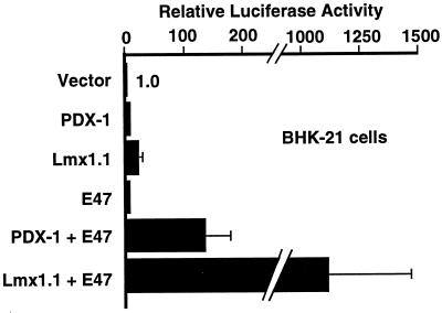
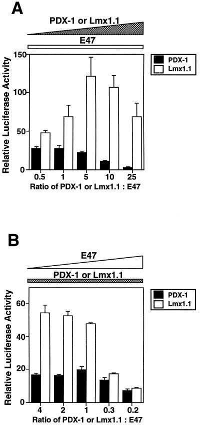
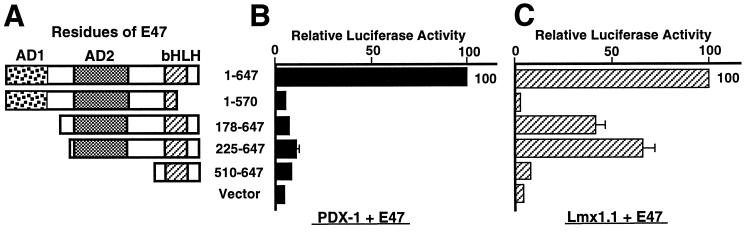
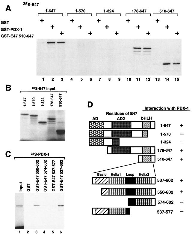
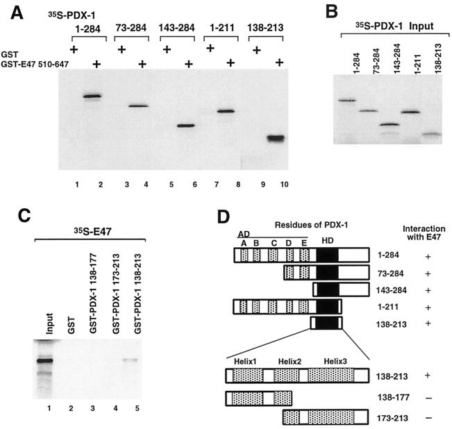
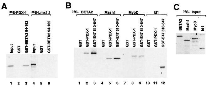
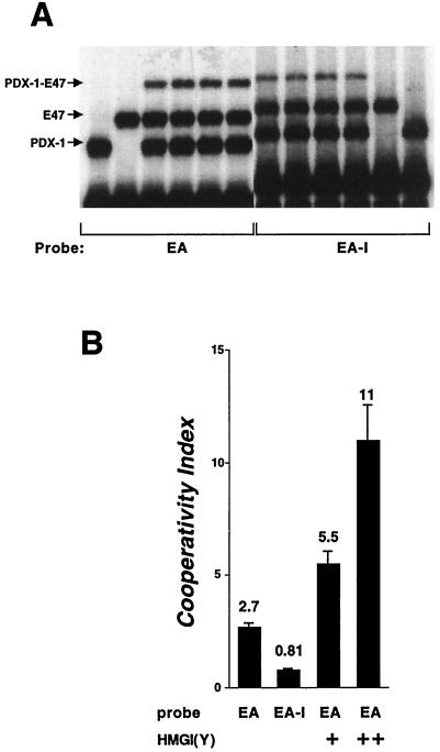
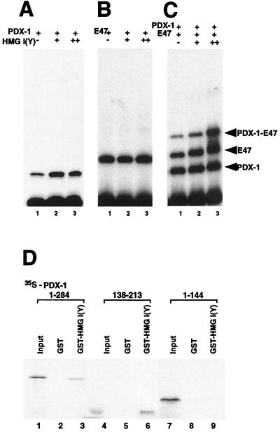
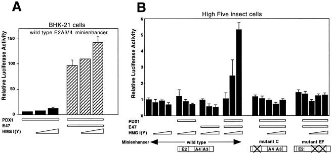
Activation of insulin gene transcription specifically in the pancreatic beta cells depends on multiple nuclear proteins that interact with each other and with sequences on the insulin gene promoter to build a transcriptional activation complex. The homeodomain protein PDX-1 exemplifies such interactions by binding to the A3/4 region of the rat insulin I promoter and activating insulin gene transcription by cooperating with the basic-helix-loop-helix (bHLH) protein E47/Pan1, which binds to the adjacent E2 site. The present study provides evidence that the homeodomain of PDX-1 acts as a protein-protein interaction domain to recruit multiple proteins, including E47/Pan1, BETA2/NeuroD1, and high-mobility group protein I(Y), to an activation complex on the E2A3/4 minienhancer. The transcriptional activity of this complex results from the clustering of multiple activation domains capable of interacting with coactivators and the basal transcriptional machinery. These interactions are not common to all homeodomain proteins: the LIM homeodomain protein Lmx1.1 can also activate the E2A3/4 minienhancer in cooperation with E47/Pan1 but does so through different interactions. Cooperation between Lmx1.1 and E47/Pan1 results not only in the aggregation of multiple activation domains but also in the unmasking of a potent activation domain on E47/Pan1 that is normally silent in non-beta cells. While more than one activation complex may be capable of activating insulin gene transcription through the E2A3/4 minienhancer, each is dependent on multiple specific interactions among a unique set of nuclear proteins.


Figures

FIG. 1
FIG. 1
Lmx1.1 and E47/Pan1 give greater transcriptional synergy than PDX-1 and E47/Pan1. BHK-21 cells were transfected with a reporter plasmid (pFOXluc.prl.5FF1) containing five copies of the insulin E2A3/4 minienhancer linked to the minimal prolactin promoter driving luciferase gene expression and with expression plasmids containing the CMV promoter driving expression of the cDNAs shown. Luciferase activity in the cells transfected with the reporter plasmid and the parent expression plasmid without insert is set to 1.0.
FIG. 2
FIG. 2
Excess PDX-1 decreases synergy with E47/Pan1, and excess E47/Pan1 decreases synergy with Lmx1.1. (A) The pFOXluc.prl.5FF1 reporter plasmid was cotransfected with 0.02 μg of the E47/Pan1 expression plasmids and increasing amounts (0.01, 0.02, 0.1, 0.2, and 0.5 μg) of the PDX-1 or Lmx1.1 expression plasmid into BHK-21 cells. Luciferase activity in the cells transfected with the reporter plasmid and the E47/Pan1 expression plasmid alone is set at 1.0. (B) The reporter plasmid was cotransfected with 0.2 μg of the PDX-1 or Lmx1.1 expression plasmid and increasing amounts (0.05, 0.1, 0.2, 0.6, and 1.0 μg) of the E47/Pan1 expression plasmid into BHK-21 cells. The luciferase activity in the cells transfected with the reporter and the PDX-1 or Lmx1.1 expression plasmid alone is set at 1.0.
FIG. 3
FIG. 3
PDX-1 and Lmx1.1 cooperate with different activation domains of E47/Pan1. The pFOXluc.prl.5FF1 reporter plasmid was cotransfected with a plasmid expressing the wild-type or truncated E47/Pan1 cDNA and a plasmid expressing either the PDX-1 or the Lmx1.1 cDNA into BHK-21 cells. Luciferase activity in the cells transfected with the reporter, the homeobox cDNA expression plasmid, and the wild-type E47/Pan1 (amino acids 1 to 647) expression plasmid is set at 100. (A) Diagram showing the E47/Pan1 deletions used. (B) Results for cells transfected with the E47/Pan1 and PDX-1 expression plasmids. (C) Results for cells transfected with the E47/Pan1 and Lmx1.1 expression plasmids.
FIG. 4
FIG. 4
PDX-1 physically interacts with the HLH domain of E47/Pan1 in vitro. (A) In vitro-translated, 35S-labeled E47/Pan1 (wild type and deletion mutants) was incubated with GST–PDX-1 fusion protein immobilized on glutathione-Sepharose beads, and bound proteins were resolved on SDS-PAGE followed by autoradiography. GST alone and GST fused to E47/Pan1 amino acids 510 to 647 (including the bHLH dimerization domain) were used as controls. (B) Ten percent of the 35S-labeled E47/Pan1 (wild-type and deletion mutant) proteins used in the binding assay were resolved by SDS-PAGE. (C) In vitro-translated, 35S-labeled PDX-1 protein was incubated with GST alone or with GST fused to the truncated E47/Pan1 proteins shown. Bound proteins were immobilized on glutathione-Sepharose beads and resolved by SDS-PAGE followed by autoradiography. Ten percent of the 35S-labeled PDX-1 protein used in the binding assay was loaded in lane 1. (D) Structures of the E47/Pan1 deletion mutants and summary of the results.
FIG. 5
FIG. 5
The intact PDX-1 homeodomain interacts physically with E47/Pan1. (A) In vitro-translated, 35S-labeled PDX-1 (wild type and deletion mutants) was incubated with GST-E47/Pan1 fusion protein immobilized on glutathione-Sepharose beads, and bound proteins were resolved by SDS-PAGE followed by autoradiography. GST alone was used as a control. (B) Ten percent of the 35S-labeled PDX-1 (wild-type and deletion mutant) proteins used in the binding assay were resolved by SDS-PAGE. (C) In vitro-translated, 35S-labeled E47/Pan1 protein was incubated with GST alone or with GST fused to the truncated PDX-1 proteins shown. Bound proteins were immobilized on glutathione-Sepharose beads and resolved by SDS-PAGE followed by autoradiography. Ten percent of the 35S-labeled E47/Pan1 protein used in the binding assay was loaded in lane 1. (D) Structures of the PDX-1 deletion mutants and summary of the results. AD, transcription activation domain; HD, homeodomain; A to E, conserved segments of the PDX-1 activation domain as described by Peshavaria et al. (40).
FIG. 6
FIG. 6
PDX-1 physically interacts with tissue-specific bHLH proteins. (A) In vitro-translated, 35S-labeled PDX-1 or Lmx1.1 protein was incubated with GST alone or with the GST-BETA2 bHLH domain (containing amino acids 94 to 162 of BETA2/NeuroD). Bound proteins were immobilized on glutathione-Sepharose beads and resolved by SDS-PAGE followed by autoradiography. Ten percent of the 35S-labeled PDX-1 and Lmx1.1 proteins used in the binding assay were loaded in lanes 1 and 4, respectively. (B) 35S-labeled full-length HLH proteins BETA2/NeuroD, Mash1, MyoD, and Id1 were tested as for panel A for interaction with GST–PDX-1. GST alone and GST fused to E47/Pan1 amino acids 510 to 647 (including the bHLH dimerization domain) were used as controls. (C) Ten percent of the 35S-labeled HLH proteins used for the assay were resolved by SDS-PAGE.
FIG. 7
FIG. 7
BETA2/NeuroD can form a ternary complex with the bHLH domain of E47/Pan1 and the homeodomain of PDX-1 on the E2A3/4 element. (A) The PDX-1 homeodomain (320 pg), E47/Pan1 bHLH domain (30 ng), and BETA2/NeuroD bHLH domain (3 ng) recombinant proteins were tested by EMSA for the ability to bind the 32P-labeled rat insulin E2A3/4 probe. One hundred-fold molar excess of nonlabeled E2 (lane 8) or A3/4 (lane 9) oligonucleotide was used as a specific competitor. (B) BHK-21 cells were transfected with 1 μg of the reporter as described for Fig. 1 and 0.2 μg of the expression plasmid DNAs of PDX-1, E47/Pan1, and BETA2/NeuroD as shown. Luciferase activity in the cells transfected with the reporter plasmid and the pBAT12 plasmid with no insert is set to 1.0.
FIG. 8
FIG. 8
Modest cooperative DNA binding by E47 and PDX1. (A) The homeodomain of PDX-1 (160 pg; lanes 1, 3 to 10, and 12) and the bHLH domain of E47/Pan1 (6 ng; lanes 2 to 11) were incubated with the 32P-labeled rat insulin E2A3/4 probe (EA; lanes 1 to 6) or with a similarly labeled probe containing a 15-bp insertion between the E and A binding sites (EA-I; lanes 7 to 12) and analyzed by EMSA. (B) The complexes from at least five independent EMSAs were quantified with a phosphorimager, and the cooperativity index was calculated as defined in Table 2. HMG I(Y) experiments were performed as for Fig. 9, with 37 ng (+) and 110 ng (++) of HMG I(Y). Mean ± standard error of the mean is shown.
FIG. 9
FIG. 9
HMG I(Y) interacts with the activation complex on the E2A3/4 minienhancer. (A) The homeodomain of PDX-1 (160 pg) was incubated with the 32P-labeled rat insulin E2A3/4 probe with GST-HMG I(Y) (160 ng in lane 2; 320 ng in lane 3) or GST alone (320 ng in lane 1; 160 ng in lane 2) and analyzed by EMSA. (B) The bHLH domain of E47/Pan1 (6 ng) was incubated with the 32P-labeled rat insulin E2A3/4 probe with GST-HMG I(Y) (160 ng in lane 2; 320 ng in lane 3) or GST alone (320 ng in lane 1; 160 ng in lane 2) and analyzed by EMSA. (C) The homeodomain of PDX-1 (160 pg) and bHLH domain of E47/Pan1 (6 ng) were incubated with the 32P-labeled rat insulin E2A3/4 probe with GST-HMG I(Y) (160 ng in lane 2; 320 ng in lane 3) or GST alone (320 ng in lane 1; 160 ng in lane 2) and analyzed by EMSA. (E) In vitro-translated, 35S-labeled PDX-1 protein (wild type and deletion mutants) was incubated with GST alone or with GST-HMG I(Y). Bound proteins were immobilized on glutathione-Sepharose beads and resolved by SDS-PAGE followed by autoradiography. Ten percent of the 35S-labeled PDX-1 proteins were loaded in lanes 1, 4, and 7.
FIG. 10
FIG. 10
HMG I(Y) contributes to the synergy of PDX-1 and E47/Pan1. (A) Transfections were performed in BHK-21 cells as for Fig. 1A, except for the addition of increasing amounts (0.2 and 0.4 μg) of the HMG I(Y) expression plasmid. Luciferase activity in the cells transfected with the reporter plasmid and the pBAT12 plasmid with no insert is set to 1.0. (B) High Five insect cells were transfected with the pFOXluc.prl.5FF1 reporter construct (1.0 μg) as for panel or with reporter constructs with mutations of the E2 (pFOXluc.prl.5mC1) or A3/4 (pFOXluc.prl.5mEF1) sites. Expression plasmids containing the CMV promoter driving expression of the cDNAs for PDX-1 (100 ng), E47/Pan1 (100 ng), or HMG I(Y) (20, 100, or 500 ng) were cotransfected. Luciferase activity in the cells transfected with the reporter plasmid and the parent expression plasmid without insert is set to 1.0.

References

    1. Abdulkadir S A, Casolaro V, Tai A K, Thanos D, Ono S J. High mobility group I/Y protein functions as a specific cofactor for Oct-2A: mapping of interaction domains. J Leukoc Biol. 1998;64:681–691. - PubMed
    1. Ahlgren U, Jonsson J, Edlund H. The morphogenesis of the pancreatic mesenchyme is uncoupled from that of the pancreatic epithelium in IPF1/PDX1-deficient mice. Development. 1996;122:1409–1416. - PubMed
    1. Ahlgren U, Jonsson J, Jonsson L, Simu K, Edlund H. β-Cell-specific inactivation of the mouse ipf1/Pdx1 gene results in loss of the β-cell phenotype and maturity onset diabetes. Genes Dev. 1998;12:1763–1768. - PMC - PubMed
    1. Arlotta P, Rustighi A, Mantovani F, Manfioletti G, Giancotti V, Tell G, Damante G. High mobility group I proteins interfere with the homeodomains binding to DNA. J Biol Chem. 1997;272:29904–29910. - PubMed
    1. Aronheim A, Shiran R, Rosen A, Walker M D. The E2A gene product contains two separable and functionally distinct transcription activation domains. Proc Natl Acad Sci USA. 1993;90:8063–8067. - PMC - PubMed

Publication types

MeSH terms