. 2000 Feb;20(3):947-56.
doi: 10.1128/MCB.20.3.947-956.2000.

Complex effects of naturally occurring mutations in the JAK3 pseudokinase domain: evidence for interactions between the kinase and pseudokinase domains

Affiliations

Complex effects of naturally occurring mutations in the JAK3 pseudokinase domain: evidence for interactions between the kinase and pseudokinase domains

M Chen et al. Mol Cell Biol. 2000 Feb.

Abstract

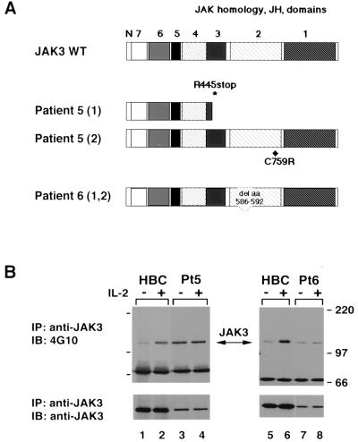
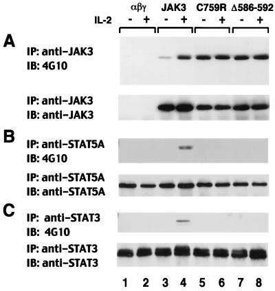
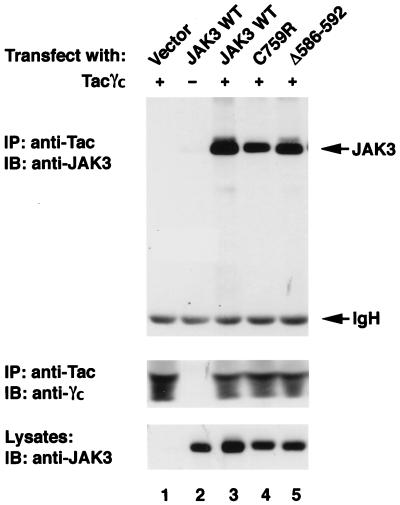
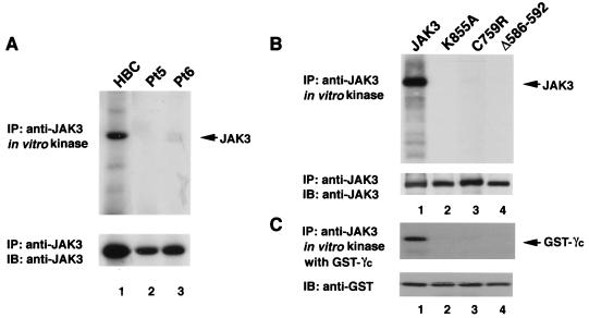
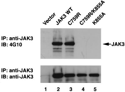
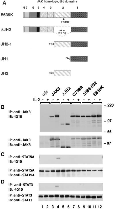
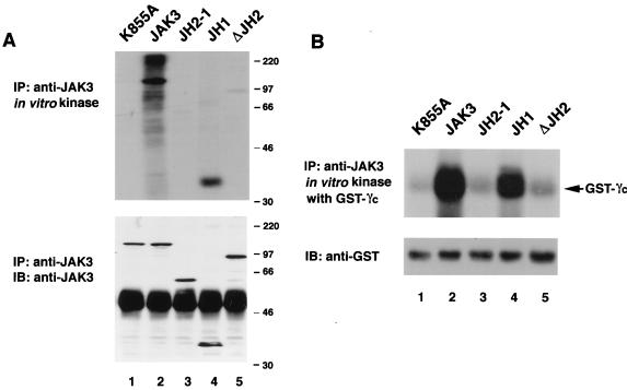
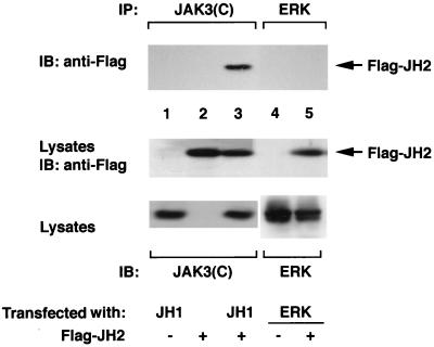
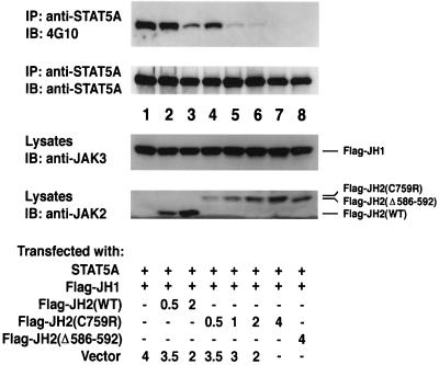
The structure of Janus kinases (JAKs) is unique among protein tyrosine kinases in having tandem, nonidentical kinase and pseudokinase domains. Despite its conservation in evolution, however, the function of the pseudokinase domain remains poorly understood. Lack of JAK3 expression results in severe combined immunodeficiency (SCID). In this study, we analyze two SCID patients with mutations in the JAK3 pseudokinase domain, which allows for protein expression but disrupts the regulation of the kinase activity. Specifically, these mutant forms of JAK3 had undetectable kinase activity in vitro but were hyperphosphorylated both in patients' Epstein-Barr virus-transformed B cells and when overexpressed in COS7 cells. Moreover, reconstitution of cells with these mutants demonstrated that, although they were constitutively phosphorylated basally, they were unable to transmit cytokine-dependent signals. Further analysis showed that the isolated catalytic domain of JAK3 was functional whereas either the addition of the pseudokinase domain or its deletion from the full-length molecule reduced catalytic activity. Through coimmunoprecipitation of the isolated pseudokinase domain with the isolated catalytic domain, we provide the first evidence that these two domains interact. Furthermore, whereas the wild-type pseudokinase domain modestly inhibited kinase domain-mediated STAT5 phosphorylation, the patient-derived mutants markedly inhibited this phosphorylation. We thus conclude that the JAK3 pseudokinase domain is essential for JAK3 function by regulating its catalytic activity and autophosphorylation. We propose a model in which this occurs via intramolecular interaction with the kinase domain and that increased inhibition of kinase activity by the pseudokinase domain likely contributes to the disease pathogenesis in these two patients.


Figures

FIG. 1
FIG. 1
JAK3 from SCID patients' cells is constitutively phosphorylated and unresponsive to IL-2 stimulation. (A) Illustration of patient alleles. Patient 5 is a compound heterozygote bearing two different alleles, one encoding a missense mutation substituting a cysteine for an arginine at codon 759 and the other encoding a premature stop codon at residue 445. Both patient 6-derived alleles encode a deletion of aa 586 to 592 in the JH2 domain of JAK3. (B) EBV-transformed B cells (1.5 × 10⁷ cells) from a healthy individual (HBC) and the JAK3-SCID patients 5 (Pt5) and 6 (Pt6) were serum starved for 4 h and then were left unstimulated (−) or were stimulated with IL-2 (1,000 U/ml) for 15 min (+); cell lysates were immunoprecipitated (IP) with anti-JAK3. Tyrosine phosphorylation was assayed by blotting (IB) with antiphosphotyrosine (4G10) (top panel). The membrane was reprobed with anti-JAK3 to confirm equal loading (bottom panel).
FIG. 2
FIG. 2
Defective IL-2 responsiveness in IL-2R-expressing fibroblasts reconstituted with patient-derived mutants. NIH 3T3 fibroblasts reconstituted with the IL-2R complex (αβγ cells) were stably transfected with cDNAs encoding wild-type JAK3 and patient 5- and 6-derived mutants C759R and Δ586-592, respectively. Serum-starved cells were left unstimulated (−) or were stimulated with IL-2 for 15 min (+); cell lysates were immunoprecipitated (IP) with anti-JAK3 (A), anti-STAT5A (B), or anti-STAT3 (C) and then subjected to immunoblotting (IB) with antiphosphotyrosine (4G10) (top panels). Membranes were reprobed with anti-JAK3 (A), anti-STAT5A (B), or anti-STAT3 (C) to confirm equal loading (bottom panels).
FIG. 3
FIG. 3
Patients' JH2 mutations do not disrupt the ability of JAK3 to bind γc. COS7 cells were transfected with 3 μg of the indicated cDNAs. Lysates were immunoprecipitated (IP) with anti-Tac and blotted with anti-JAK3 (top panel) or anti-γc (middle panel). Expression levels of various JAK3 proteins were analyzed by immunoblotting (IB) with anti-JAK3 (bottom panel). WT, wild type.
FIG. 4
FIG. 4
Patient-derived JH2 mutants lack in vitro kinase activity. (A) EBV-transformed B cells (1.5 × 10⁷ cells) from HBC (lanes 1) and patients 5 (Pt5) and 6 (Pt6) were treated with IL-2 (15 min), lysed, immunoprecipitated (IP) with anti-JAK3, and then subjected to an in vitro kinase assay. Samples were analyzed by sodium dodecyl sulfate-polyacrylamide gel electrophoresis and transferred to a polyvinylidene difluoride membrane. Incorporated radioactive phosphate was visualized by autoradiography (top panel). Expression levels of JAK3 were determined by immunoblotting (IB) with anti-JAK3 (bottom panel). (B and C) Wild-type JAK3 (lanes 1), catalytically inactive JAK3 (K855A) (lanes 2), and patient 5- and 6-derived mutants (lanes 3, C759R; lanes 4, Δ586-592) were expressed in COS7 cells. Cell lysates were immunoprecipitated with anti-JAK3 and subjected to an in vitro kinase assay. Autophosphorylation (B) and phosphorylation of an exogenous substrate, GST-γc (C), are shown in the top panels. Membranes were probed with relevant antibody to verify equal expression and loading (bottom panels).
FIG. 5
FIG. 5
Patient 5-derived JH2 mutant C759R is hyperphosphorylated when expressed in COS7 cells. COS7 cells were transfected with 3 μg of the indicated cDNAs. The cells were lysed, immunoprecipitated (IP) with anti-JAK3, and blotted (IB) with antiphosphotyrosine antibody 4G10 (top panel). The membrane was then stripped and reprobed with anti-JAK3 to show the levels of various JAK3 proteins (bottom panel).
FIG. 6
FIG. 6
The JH2 pseudokinase domain regulates JAK3 kinase activity and substrate phosphorylation. (A) Schematic representation of the JH2 mutants. E639K is a JAK3 mutant corresponding to the hyperactivating mutation reported in Drosophila Hopscotch (26). ΔJH2 contains a complete deletion of the JH2 domain, from aa 519 to aa 792. (B to D) αβγ cells expressing various forms of JAK3 were left unstimulated (−) or stimulated with IL-2 for 15 min (+); cell lysates were immunoprecipitated (IP) with anti-JAK3 (B), anti-STAT5A (C), or anti-STAT3 (D) and then subjected to immunoblotting (IB) analysis with anti-phosphotyrosine (B to D, top panels). Membranes were reprobed with anti-JAK3 (B), anti-STAT5A (C), or anti-STAT3 (D) to confirm equal loading (bottom panels).
FIG. 7
FIG. 7
The JH2 pseudokinase domain inhibits kinase activity of the isolated catalytic domain of JAK3. (A) JAK3 autophosphorylation. (B) In vitro kinase assay using GST-γc as the exogenous substrate. COS7 cells were transfected with 3 μg of the indicated cDNAs. Lysates were immunoprecipitated (IP) with anti-JAK3 and then subjected to an in vitro kinase assay in the absence (A) or presence (B) of GST-γc. Incorporated radioactive phosphate was visualized by autoradiography (top panels). Immunoblotting (IB) with anti-JAK3 (A) or anti-GST (B) was performed to confirm equal loading and expression.
FIG. 8
FIG. 8
Interaction between the JH2 and JH1 domains of JAK3. COS7 cells were transfected with 3 μg of the indicated cDNAs. Lysates were immunoprecipitated (IP) with an antiserum against the C terminus of JAK3 (lanes 1 to 3) or anti-ERK (lanes 4 and 5) and subjected to immunoblotting (IB) with anti-Flag. The expression levels of Flag-tagged JH2 (middle panel) and JH1 and ERK2 (bottom panel) were analyzed by immunoblotting with the indicated antibodies.
FIG. 9
FIG. 9
Overexpression of the patient-derived JH2 domain inhibits JH1-mediated STAT5A phosphorylation to a greater extent than that of the wild type. COS7 cells were cotransfected with STAT5A (0.5 μg) and Flag-JH1 (0.5 μg) together with the indicated amounts of various JH2 constructs and the parental vector (total amount of transfected DNA was 5 μg in all cases). Lysates were immunoprecipitated (IP) with anti-STAT5A and blotted (IB) with anti-phosphotyrosine (4G10) (first panel) and anti-STAT5A (second panel). The expression levels of Flag-JH1 and various JH2 proteins were analyzed by immunoblotting with anti-JAK3 (third panel) and an antibody that recognizes the JH2 domains of JAK2 and JAK3 (bottom panel).

References

    1. Boussiotis V A, Barber D L, Nakarai T, Freeman G J, Gribben J G, Bernstein G M, D'Andrea A D, Ritz J, Nadler L M. Prevention of T cell anergy by signaling through the gamma c chain of the IL-2 receptor. Science. 1994;266:1039–1042. - PubMed
    1. Cacalano N A, Migone T S, Bazan F, Hanson E P, Chen M, Candotti F, O'Shea J J, Johnston J A. Autosomal SCID caused by a point mutation in the amino-terminus of JAK3: mapping of the JAK3-receptor interaction domain. EMBO J. 1999;18:1549–1558. - PMC - PubMed
    1. Candotti F, O'Shea J J, Villa A. Severe combined immune deficiencies due to defects of the common gamma chain-JAK3 signaling pathway. Springer Semin Immunopathol. 1998;19:401–415. - PubMed
    1. Candotti F, Oakes S A, Johnston J A, Giliani S, Schumacher R F, Mella P, Fiorini M, Ugazio A G, Badolato R, Notarangelo L D, Bozzi F, Macchi P, Strina D, Vezzoni P, Blaese R M, O'Shea J J, Villa A. Structural and functional basis for JAK3-deficient severe combined immunodeficiency. Blood. 1997;90:3996–4003. - PubMed
    1. Chen M, Cheng A, Chen Y Q, Hymel A, Hanson E P, Kimmel L, Minami Y, Taniguchi T, Changelian P S, O'Shea J J. The amino terminus of JAK3 is necessary and sufficient for binding to the common gamma chain and confers the ability to transmit interleukin 2-mediated signals. Proc Natl Acad Sci USA. 1997;94:6910–6915. - PMC - PubMed

Publication types

MeSH terms

Grants and funding

LinkOut - more resources