. 2000 Feb;20(3):1044-54.
doi: 10.1128/MCB.20.3.1044-1054.2000.

Death-associated protein kinase-related protein 1, a novel serine/threonine kinase involved in apoptosis

Affiliations

Death-associated protein kinase-related protein 1, a novel serine/threonine kinase involved in apoptosis

B Inbal et al. Mol Cell Biol. 2000 Feb.

Abstract

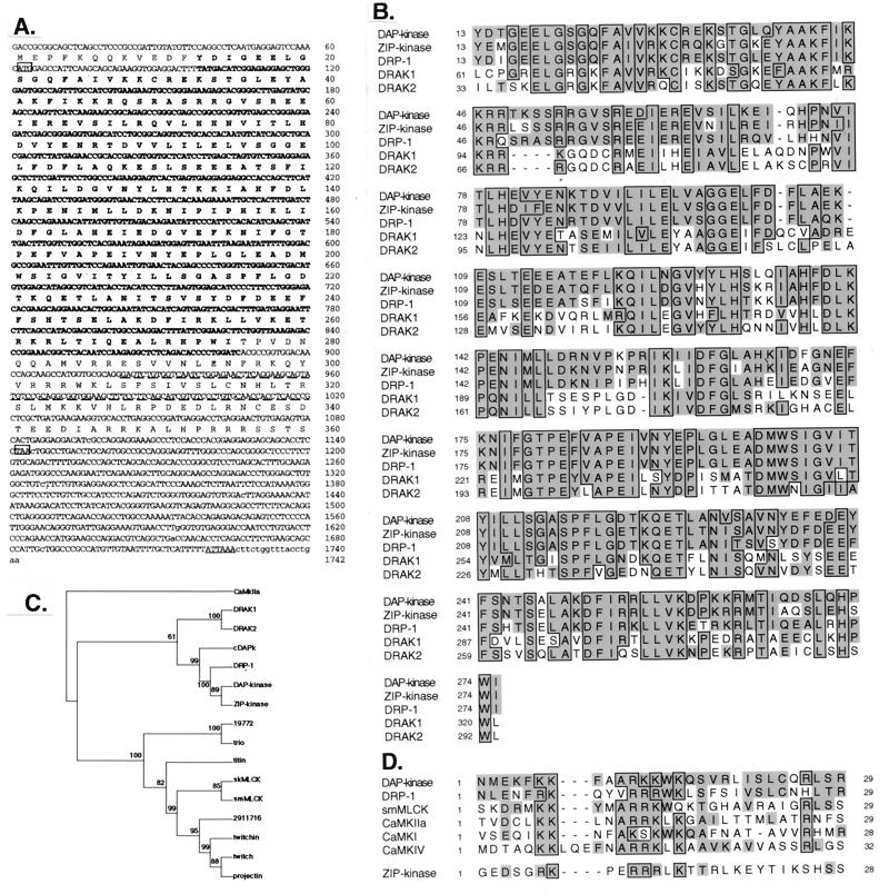
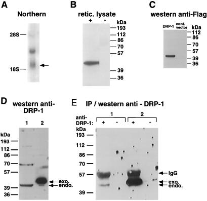
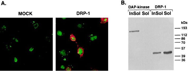
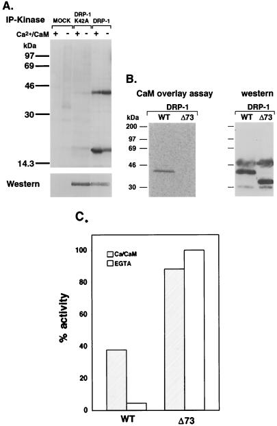
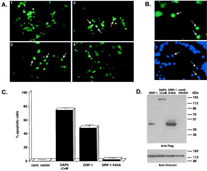
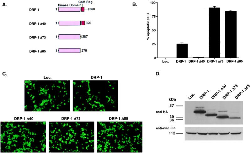
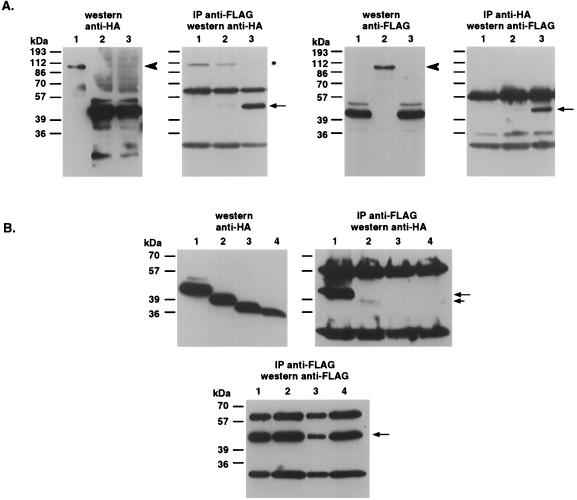
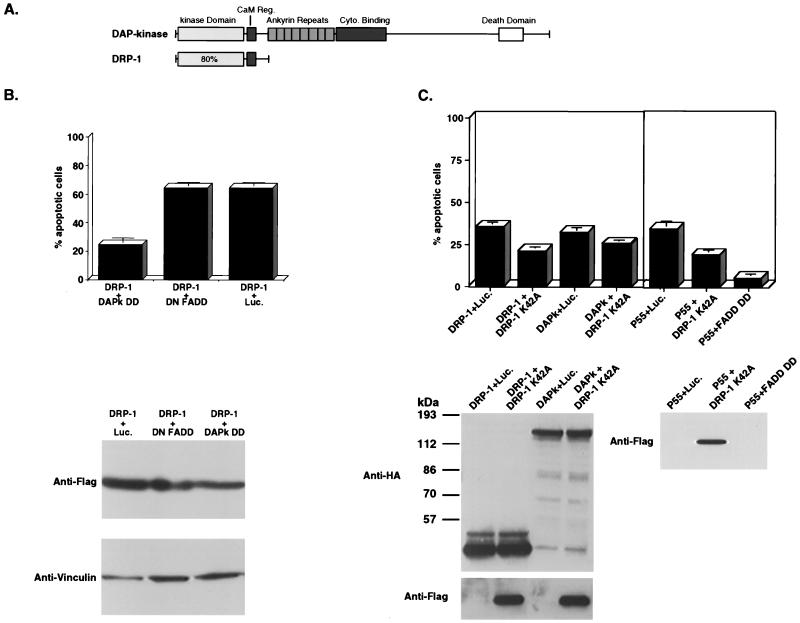
In this study we describe the identification and structure-function analysis of a novel death-associated protein (DAP) kinase-related protein, DRP-1. DRP-1 is a 42-kDa Ca(2+)/calmodulin (CaM)-regulated serine threonine kinase which shows high degree of homology to DAP kinase. The region of homology spans the catalytic domain and the CaM-regulatory region, whereas the remaining C-terminal part of the protein differs completely from DAP kinase and displays no homology to any known protein. The catalytic domain is also homologous to the recently identified ZIP kinase and to a lesser extent to the catalytic domains of DRAK1 and -2. Thus, DAP kinase DRP-1, ZIP kinase, and DRAK1/2 together form a novel subfamily of serine/threonine kinases. DRP-1 is localized to the cytoplasm, as shown by immunostaining and cellular fractionation assays. It binds to CaM, undergoes autophosphorylation, and phosphorylates an exogenous substrate, the myosin light chain, in a Ca(2+)/CaM-dependent manner. The truncated protein, deleted of the CaM-regulatory domain, was converted into a constitutively active kinase. Ectopically expressed DRP-1 induced apoptosis in various types of cells. Cell killing by DRP-1 was dependent on two features: the status of the catalytic activity, and the presence of the C-terminal 40 amino acids shown to be required for self-dimerization of the kinase. Interestingly, further deletion of the CaM-regulatory region could override the indispensable role of the C-terminal tail in apoptosis and generated a "superkiller" mutant. A dominant negative fragment of DAP kinase encompassing the death domain was found to block apoptosis induced by DRP-1. Conversely, a catalytically inactive mutant of DRP-1, which functioned in a dominant negative manner, was significantly less effective in blocking cell death induced by DAP kinase. Possible functional connections between DAP kinase and DRP-1 are discussed.


Figures

FIG. 1
FIG. 1
Sequence of the DRP-1 cDNA clone and alignments to related kinases. (A) Nucleotide and deduced amino acid sequences of human DRP-1. Initiation (ATG) and stop (TAA) codons are boxed. A polyadenylation signal (ATTAAA) is underlined. The kinase domain and the CaM-regulatory regions are in bold and underlined by dashes, respectively. (B) Multiple sequence alignment of the serine/threonine kinase domains of the related proteins DAP kinase, ZIP kinase, DRP-1, and DRAK1 and -2. Alignment was done as described by Hanks and Quinn (14). Identical amino acids are boxed; homologous amino acids according to PAM250 matrix are shown in grey. (C) Phylogenic rooted neighbor-joining tree of the 16 catalytic domains belonging to proteins closely related to DAP kinase. Numbers shown are bootstrap values. Confidence values lower than 50% are considered unreliable. CaMKIIa was used as a representative of other CaM kinases and was outgrouped to root the tree. smMLCK and skMLCK, smooth muscle and skeletal MLCK, respectively. (D) Multiple sequence alignment of the CaM-regulatory regions of DAP kinase, DRP-1, smMLCK, CaMKIIa, CaMKI, and CaMKIV. Alignment was done manually, keeping the conserved (boxed) regions aligned to each other. The corresponding region of ZIP kinase which does not contain homology to DAP kinase and DRP-1 CaM-regulatory regions is given at the bottom.
FIG. 2
FIG. 2
mRNA and protein expression of DRP-1. (A) Northern blot analysis of DRP-1 mRNA. A 580-bp 3′ fragment from the full-length cDNA of DRP-1 was used to probe poly(A)+ RNA prepared from MCF-7 cells. (B) In vitro translation of DRP-1. In vitro-transcribed DRP-1 mRNA was programmed in reticulocyte lysate. The translated protein, 42 kDa in size, is shown. (C) Protein expression of recombinant DRP-1 in HeLa cells. FLAG-tagged DRP-1 cloned in the pCDNA3 vector was transfected into HeLa cells; 24 h following transfection, cells were harvested and lysed. Extracted proteins were separated by SDS-PAGE and then immunoblotted with anti-FLAG antibodies. A protein band at ∼42 kDa is shown. (D and E) Expression of endogenous (endo.) DRP-1 protein. (D) Western analysis in which 100 μg of protein lysates of MCF-7 cells (1) and 30 μg of protein lysates of 293 cells transfected by DRP-1-FLAG (2) were separated by SDS-PAGE and immunoblotted with 9% anti-DRP-1 serum. exo., exogenous. (E) IP/Western analysis in which 3 mg of protein lysates of MCF-7 cells (1) or 300 μg of protein lysates of 293 cells transfected by DRP-1-FLAG (2) was immunoprecipitated overnight with (+) or without (−) 50 μl of anti DRP-1 serum. The proteins were separated by SDS-PAGE and immunoblotted with 9% anti-DRP-1 serum. Arrows indicate positions of the endogenous and exogenous DRP-1 and the position of immunoglobulin G (IgG) heavy-chain protein.
FIG. 3
FIG. 3
Intracellular localization of DRP-1 in COS-7 cells. (A) COS-7 cells were transfected by a FLAG-tagged DRP-1 cloned in pCDNA3 vector, fixed and permeabilized in 1% formaldehyde, and treated with methanol-acetone. Fixed cells were then reacted with Oligreen for nuclear staining (green) and with anti-FLAG antibodies for DRP-1 detection (red). Cells were visualized under a fluorescence microscope. (B) Detergent extraction of COS-7 cells. COS-7 cells were transfected with a pCDNA3 vector expressing either FLAG-tagged DRP-1 or DAP kinase. The cells were then extracted with 0.5% Triton X-100 to form soluble fractions (Sol) and insoluble fractions (InSol) as described in Materials and Methods. The protein extracts were separated by 10% SDS-PAGE (10% gel) and blotted onto a nitrocellulose membrane. The membrane was reacted with anti-FLAG antibodies.
FIG. 4
FIG. 4
In vitro kinase activity and CaM binding of DRP-1. (A) DRP-1-FLAG and DRP-1-FLAG mutant (K42A) proteins were assayed in vitro for kinase activity in the presence or absence of Ca2+/CaM as described in Materials and Methods. The proteins were resolved by SDS-PAGE on an 11% gel and blotted to a nitrocellulose membrane. Top, autophosphorylation of DRP-1 (42 kDa) and MLC phosphorylation (17 kDa), respectively, after exposure to X-ray film; bottom, DRP-1 proteins after incubation of the same blot with anti-FLAG antibodies and ECL detection. (B) Cell lysates were prepared from 293 cells transfected with wild-type (WT) DRP-1-HA or DRP-1-HA Δ73 mutant and separated by PAGE-SDS on a 12% gel. Blotted proteins were reacted with recombinant 35S-labeled CaM (left); exogenous proteins were detected with anti-HA antibodies in a standard Western blot procedure (right). (C) Graph showing the relative amount of MLC phosphorylation by wild-type DRP-1 or DRP-1 Δ73 mutant in the presence or absence of Ca2+/CaM. Phosphorylation levels were determined by phosphorimaging of 32P-labeled MLC bands. Values were normalized according to the DRP-1 recombinant protein levels used in each phosphorylation assay. Phosphorylation of MLC by DRP-1 Δ73 in the absence of Ca2+/CaM was taken as the maximal phosphorylation activity.
FIG. 5
FIG. 5
Ectopic expression of DRP-1 induces cell death. (A) 293 cells (105 cells/well) were cotransfected with FLAG-tagged wild-type DRP-1 or K42A mutant DRP-1 (1.5 μg/well) and GFP (0.5 μg/well). GFP-positive cells were visualized under a fluorescence microscope and scored for appearance of apoptotic morphology 24 h after transfection. Apoptotic cells are indicated by arrows. Images 1 to 4 correspond to 293 cells transfected by pCDNA3-luciferase (negative control), pCDNA3-ΔCaM DAP kinase (positive control), pCDNA3-DRP-1, and pCDNA3-DRP-1 K42A. (B) Nuclear staining of DRP-1-transfected cells. Top, GFP staining of 293 cells transfected by DRP-1 Δ73 mutant (1.5 μg/well); bottom, Hoechst nuclear staining of the same cells. Pictures were taken 60 h posttransfection. Solid arrows, cells with condensed and fragmented chromatin; dashed arrows, cells with condensed chromatin. (C) Scores of apoptotic cells. Graphs show the percentage of apoptotic cells resulting from the above-mentioned transfections (mean ± standard deviation calculated from triplicates of 100 cells each). The scores were taken from the same experiment as shown in panel A. This experiment was repeated six times with reproducible results. (D) DRP-1 protein expression in transfected 293 cells. Proteins extracted from the transfected cells were separated by SDS-PAGE on a 10% gel and blotted to a nitrocellulose membrane. The blot was reacted with anti-FLAG antibodies for DRP-1 detection and antivinculin antibodies (dilution of 1:300; Sigma) to quantitate the loaded protein amounts. The proteins were prepared from the same experiment as shown in panel A.
FIG. 6
FIG. 6
Ectopic expression of DRP-1 deletion mutants. (A) Schematic representation of DRP-1 deletion mutants. (B) Scores of apoptotic cells. Graphs show the percentage of apoptotic cells resulting from cotransfections of 293 cells with 1.2 μg of HA-tagged wild-type DRP-1 or various deletion mutants of DRP-1 (mean ± standard deviation calculated from triplicates of 100 cells each). This experiment was repeated three times with reproducible results. (C) Pictures were taken from the experiment described above, corresponding to 293 cells transfected with pCDNA3-luciferase (Luc.; negative control), pCDNA3-DRP-1, pCDNA3-DRP-1 Δ40, pCDNA3-DRP-1 Δ73, and pCDNA3-DRP-1 Δ85. (D) DRP-1 protein expression in transfected 293 cells. Proteins extracted from the transfected cells were separated by SDS-PAGE on a 10% gel and blotted to nitrocellulose membrane. The blot was reacted with anti-HA antibodies for DRP-1 detection and antivinculin antibodies to quantitate the loaded protein amounts. The proteins were prepared from the same experiment as shown in panel B.
FIG. 7
FIG. 7
DRP-1 undergoes homodimerization via its C-terminal tail. (A) Wild-type DRP-1 undergoes specific homodimerization. 293 cells growing in 90-mm-diameter plates were cotransfected with the following constructs: lane 1, DRP-1-FLAG (5 μg) plus RFX1-ΔSmaI-HA (20 μg; used as an intrinsic control to rule out nonspecific binding of DRP-1-FLAG to HA beads or to an irrelevant gene); lane 2, RFX-ΔSmaI-FLAG plus DRP-1-HA (control to rule out nonspecific attachment of DRP-1-HA to FLAG beads or to an irrelevant gene); lane 3, DRP-1-FLAG plus DRP-1-HA. Cell extracts were immunoprecipitated with either anti-FLAG antibodies (left) or anti-HA antibodies (right). The total levels of transfected proteins are shown on immunoblots. (B) Truncation of C-terminal 40 amino acids of DRP-1 abolishes its homodimerization. Lanes 1 to 4 correspond to cotransfections (5 μg of each construct per 90-mm-diameter plate) with DRP-1-FLAG plus DRP-1-HA, DRP-1-FLAG plus DRP-1-Δ40-HA, DRP-1-FLAG plus DRP-1-Δ73-HA, and DRP-1-FLAG plus DRP-1-Δ85-HA, respectively. The lower panel quantitates the immunoprecipitation efficiency of DRP-1-FLAG with the anti-FLAG antibodies. Solid arrows, immunoprecipitated DRP-1; short arrow, immunoprecipitated DRP-1 Δ40; arrowhead, RFX-ΔSmaI; asterisk, nonspecific band.
FIG. 8
FIG. 8
Death protection assays. (A) Schematic representation of DRP-1 and DAP kinase. (B) DAP kinase death domain protects from DRP-1-induced cell death. Top, 293 cells (105 cells/well) were cotransfected with FLAG-tagged wild-type DRP-1 and GFP (0.5 μg/well) as described for Fig. 5. To each transfection the indicated plasmid (DAPk DD, DN FADD, or luciferase [Luc.] in pCDNA3) was also added (0.5 μg/well). Scores are percentages of apoptotic cells given as the mean ± standard deviation and calculated from triplicates of 100 cells each. This experiment was repeated three times with reproducible results. Bottom, DRP-1 protein expression in transfected 293 cells. Proteins extracted from the transfected cells were separated by SDS-PAGE on a 10% gel and blotted to a nitrocellulose membrane. The blot was reacted with anti-FLAG antibodies for DRP-1 detection and antivinculin antibodies to quantitate the loaded protein amounts. The proteins were prepared from the same experiment shown in the top part of panel B. (C) DRP-1 K42A mutant protects from DRP-1 and p55 TNFR-induced cell deaths. Top, 293 cells (105 cells/well) were cotransfected with HA-tagged wild-type DRP-1 or HA-tagged wild-type DAP kinase (1.3 μg/well; left) or p55 TNFR (0.1 μg/well; right) and GFP (0.5 μg/well) as described for Fig. 5. To each transfection was added: the indicated plasmid DRP-1-FLAG K42A or luciferase in pCDNA3 (0.75 μg/well; left) or DRP-1-FLAG K42A, FADD DD, or luciferase in pCDNA3 (1.6 μg/well; right). Scores are the percentage of apoptotic cells given as the mean ± standard deviation and calculated from triplicates of 100 cells each. This experiment was repeated three times with reproducible results. Bottom, DRP-1-HA, HA-DAP kinase, and DRP-1-FLAG K42A protein expression in 293 transfected cells. The proteins were prepared from the same experiment as shown in the upper part of panel C.

References

    1. Anderson P. Kinase cascades regulating entry into apoptosis. Microbiol Mol Biol Rev. 1997;61:33–46. - PMC - PubMed
    1. Basu S, Kolesnick R. Stress signals for apoptosis: ceramide and c-Jun kinase. Oncogene. 1998;17:3277–3285. - PubMed
    1. Bokoch G M. Caspase-mediated activation of PAK2 during apoptosis: proteolytic kinase activation as a general mechanism of apoptotic signal transduction? Cell Death Differ. 1998;5:637–645. - PubMed
    1. Cardone M H, Salvesen G S, Widmann C, Johnson G, Frisch S M. The regulation of anoikis: MEKK-1 activation requires cleavage by caspases. Cell. 1997;90:315–323. - PubMed
    1. Cardone M H, Roy N, Stennicke H R, Salvesen G S, Franke T F, Stanbridge E, Frisch S, Reed J C. Regulation of cell death protease caspase-9 by phosphorylation. Science. 1998;282:1318–1321. - PubMed

Publication types

MeSH terms

Substances