. 2000 Jan;182(2):264-71.
doi: 10.1128/JB.182.2.264-271.2000.

Mutational analysis of a role for salicylic acid in iron metabolism of Mycobacterium smegmatis

Affiliations

Mutational analysis of a role for salicylic acid in iron metabolism of Mycobacterium smegmatis

T Adilakshmi et al. J Bacteriol. 2000 Jan.

Abstract

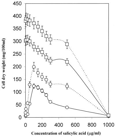
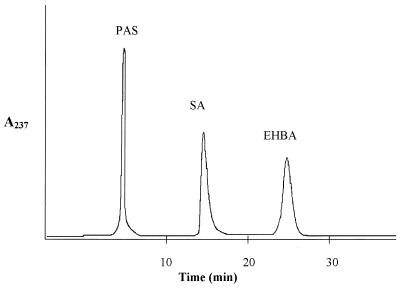
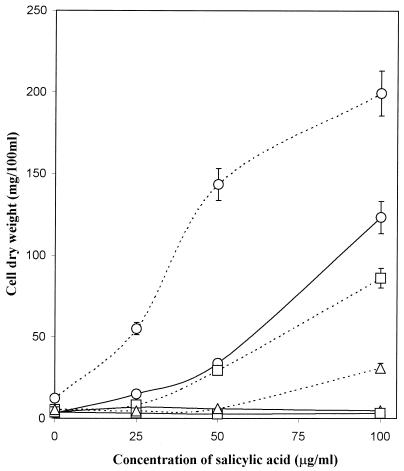
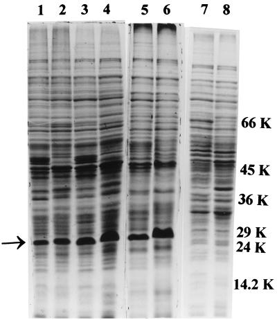
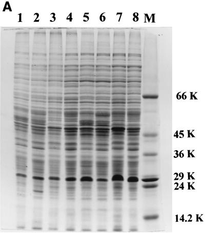
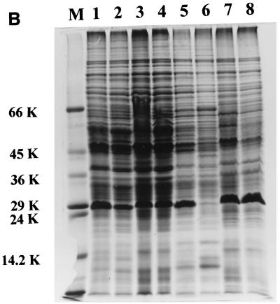
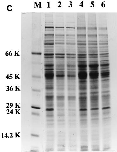
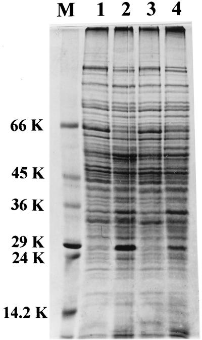
The role of salicylic acid in iron metabolism was examined in two wild-type strains (mc(2)155 and NCIMB 8548) and three mutant strains (mc(2)1292 [lacking exochelin], SM3 [lacking iron-dependent repressor protein IdeR] and S99 [a salicylate-requiring auxotroph derived in this study]) of Mycobacterium smegmatis. Synthesis of salicylate in SM3 was derepressed even in the presence of iron, as was synthesis of the siderophores exochelin, mycobactin, and carboxymycobactin. S99 was dependent on salicylate for growth and failed to grow with the three ferrisiderophores, suggesting that salicylate fulfills an additional function(s) other than being a precursor of mycobactin and carboxymycobactin. Salicylic acid at 100 microgram/ml repressed the formation of a 29-kDa cell envelope protein (putative exochelin receptor protein) in S99 grown both iron deficiently and iron sufficiently. In contrast, synthesis of this protein was affected only under iron-limited conditions in the parent strain, mc(2)155, and remained unaltered in SM3, suggesting an interaction between the IdeR protein and salicylate. Thus, salicylate may also function as a signal molecule for recognition of cellular iron status. Growth of all strains and mutants with p-aminosalicylate (PAS) at 100 microgram/ml increased salicylate accumulation between three- and eightfold under both iron-limited and iron-sufficient growth conditions and decreased mycobactin accumulation by 40 to 80% but increased carboxymycobactin accumulation by 50 to 55%. Thus, although PAS inhibited salicylate conversion to mycobactin, presumptively by blocking salicylate AMP kinase, PAS also interferes with the additional functions of salicylate, as its effect was heightened in S99 when the salicylate concentration was minimal.


Figures

FIG. 1
FIG. 1
Responses of wild-type M. smegmatis mc2155 (□) and salicylate-requiring mutant S99 (○) to salicylate under iron-insufficient (———) and iron-sufficient (–––) conditions.
FIG. 2
FIG. 2
Separation of salicylic acid (SA) and PAS by HPLC with ethyl 4-hydroxybenzoic acid (EHBA) as the internal standard. Eluants were monitored by measuring A237.
FIG. 3
FIG. 3
Influence of exogenous salicylate on the growth of S99 in the absence of PAS (○) and in the presence of 1 (□) or 5 (▵) μg of PAS per ml under iron-deficient (———) and iron-sufficient (–––) conditions.
FIG. 4
FIG. 4
SDS-PAGE profile of cell envelope proteins of M. smegmatis mutant SM3 (lanes 1 and 2), parent mc2155 (lanes 3 and 4), mutant mc21292 (lanes 5 and 6), and mutant S99 (lanes 7 and 8) under iron-deficient and -sufficient conditions, respectively. The arrow indicates the 29-kDa iron-regulated envelope protein. Molecular masses (kilodaltons) are shown on the right.
FIG. 5
FIG. 5
SDS-PAGE profile of cell envelope proteins of M. smegmatis. The effect of exogenous salicylic acid on the 29-kDa protein in the SM3 mutant (lanes 1 to 4) and the parent strain mc2155 (lanes 5 to 8) is shown. Lanes: 1 and 5, low iron; 2 and 6, low iron plus salicylic acid; 3 and 7, high iron; 4 and 8, high iron plus salicylic acid. Salicylic acid was added at 25 (A) and 100 (B) μg/ml. (C) Wild-type M. smegmatis NCIMB 8548 grown with different concentrations of salicylic acid under iron-deficient (lanes 1 to 3) and iron-sufficient (lanes 4 to 6) conditions. Lanes: 1 and 4, no salicylic acid; 2 and 5, 50 μg of salicylic acid/ml; 3 and 6, 100 μg of salicylic acid/ml. Lanes M contained marker proteins whose molecular masses (kilodaltons) are shown beside the panels.
FIG. 5
FIG. 5
SDS-PAGE profile of cell envelope proteins of M. smegmatis. The effect of exogenous salicylic acid on the 29-kDa protein in the SM3 mutant (lanes 1 to 4) and the parent strain mc2155 (lanes 5 to 8) is shown. Lanes: 1 and 5, low iron; 2 and 6, low iron plus salicylic acid; 3 and 7, high iron; 4 and 8, high iron plus salicylic acid. Salicylic acid was added at 25 (A) and 100 (B) μg/ml. (C) Wild-type M. smegmatis NCIMB 8548 grown with different concentrations of salicylic acid under iron-deficient (lanes 1 to 3) and iron-sufficient (lanes 4 to 6) conditions. Lanes: 1 and 4, no salicylic acid; 2 and 5, 50 μg of salicylic acid/ml; 3 and 6, 100 μg of salicylic acid/ml. Lanes M contained marker proteins whose molecular masses (kilodaltons) are shown beside the panels.
FIG. 6
FIG. 6
SDS-PAGE profile of cell envelope proteins of parent strain mc2155 grown with 100 μg of PAS ml−1 under low- and high-iron conditions in the absence of salicylic acid (lanes 1 and 2) and in the presence of salicylic acid (lanes 3 and 4). Molecular mass marker (lane M) sizes are shown on the left in kilodaltons.

References

    1. Barclay R, Wheeler P R. Metabolism of mycobacteria in tissues. In: Ratledge C, Stanford J L, Grange J M, editors. The biology of the mycobacteria. Vol. 3. London, United Kingdom: Academic Press Ltd.; 1989. pp. 137–196.
    1. Barclay R, Ratledge C. Iron-binding compounds of Mycobacterium avium, M. intracellulare, M. scrofulaceum, and mycobactin-dependent M. paratuberculosis and M. avium. J Bacteriol. 1983;153:1138–1146. - PMC - PubMed
    1. Brown K A, Ratledge C. The effect of p-aminosalicylic acid on iron transport and assimilation in mycobacteria. Biochim Biophys Acta. 1975;385:207–220. - PubMed
    1. Calder K M, Horwitz M A. Identification of iron-regulated proteins of Mycobacterium tuberculosis and cloning of tandem genes encoding a low iron-induced protein and a metal transporting ATPase with similarities to two-component metal transport systems. Microb Pathog. 1998;24:133–143. - PubMed
    1. Cohen S P, Levy S B, Foulds J, Rosner J L. Salicylate induction of antibiotic resistance in Escherichia coli: activation of the mar operon and a mar-independent pathway. J Bacteriol. 1993;175:7856–7862. - PMC - PubMed

Publication types

MeSH terms