

Review
F-actin bundles are derivatives of microvilli: What does this tell us about how bundles might form?
Affiliations
- PMID: 10629213
- PMCID: PMC2156212
Item in Clipboard
Review
F-actin bundles are derivatives of microvilli: What does this tell us about how bundles might form?
J Cell Biol.
.
No abstract available
Figures
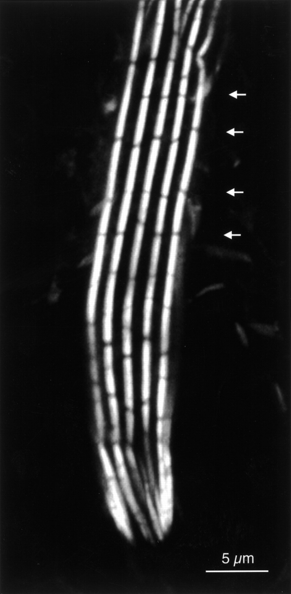
The actin bundles in a bristle of Drosophila were stained with fluorescent phalloidin and viewed with a confocal microscope. Each of the five bundles viewed in this plane is made up of smaller units or modules. Of interest is that the modules in adjacent bundles are in transverse register (see arrows). From Tilney et al., 1996.
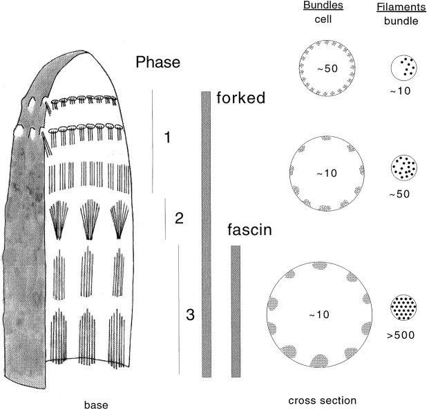
Drawing depicting the top of a Drosophila bristle at an early stage in its elongation. In stage 1 actin filaments are initiated on the cytoplasmic side of the plasma membrane in tiny pimple-like projections that resemble the tips of microvilli. Thin cross sections show that there are approximately 50 tiny bundles of roughly 10 filaments/bundle distributed evenly around the circumference of the plasma membrane. In stage 2 the tiny bundles aggregate into ∼10 large bundles each containing ∼50 filaments. The cross-linker forked is present in both stage 1 and 2. In stage 3 the cross-linker fascin enters these forked containing bundles to form along with forked hexagonally packed maximally cross-linked bundles. Additional filaments are added to the bundle and cross-linked into place. The apical end of each module is pointed, the basal end flat.
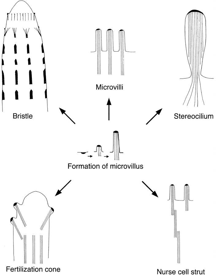
Examples of cross-linked bundles of actin filaments in a wide variety of cells from different organisms. All appear derived from the archetypal bundle, the microvillus. These include the bristle of Drosophila, the brush border of intestinal epithelial cell, the stereocilium of hair cells of the vertebrate ear, the nurse cell strut in Drosophila follicles and the fertilization cone of a sea urchin egg. An important feature of all these bundles is that the barbed ends of the actin filaments are embedded in a dense substance. Formation of a microvillus is depicted in the center and stages in the formation of a bristle are indicated. In this drawing all six parts, except the bristle (top left) are drawn to about the same size, or 1 cm equals 1 μm. The bristle is about three times smaller on this scale, or 3 mm equals ∼1 μm.
References
-
- Beckerle M.C. Spatial control of actin filament assemblylessons from Listeria . Cell. 1998;95:741–748. - PubMed
-
- Ezzell R.M., Chafel M.M., Matsudaira P.T. Differential localization of villin and fimbrin during development of the mouse visceral endoderm and intestinal epithelium. Development. 1989;106:407–419. - PubMed
Publication types
MeSH terms
Substances
Grants and funding
LinkOut - more resources
Full Text Sources
Molecular Biology Databases