. 2000 Jan;66(1):47-68.
doi: 10.1086/302722.

A physical map, including a BAC/PAC clone contig, of the Williams-Beuren syndrome--deletion region at 7q11.23

Affiliations

A physical map, including a BAC/PAC clone contig, of the Williams-Beuren syndrome--deletion region at 7q11.23

R Peoples et al. Am J Hum Genet. 2000 Jan.

Abstract

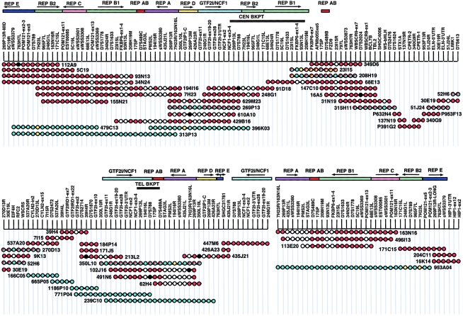
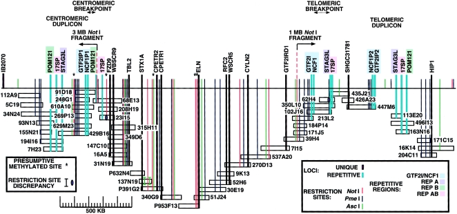
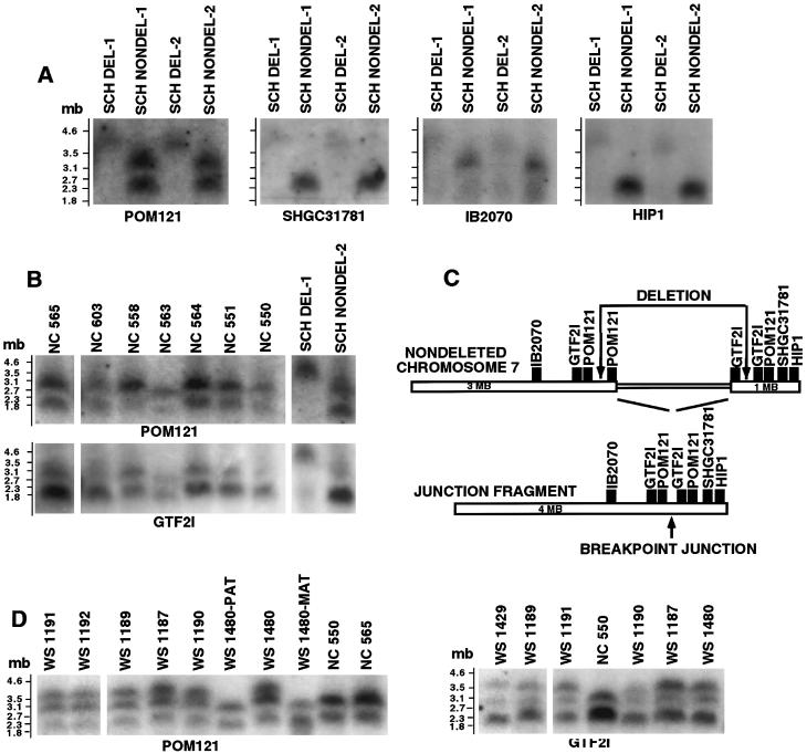
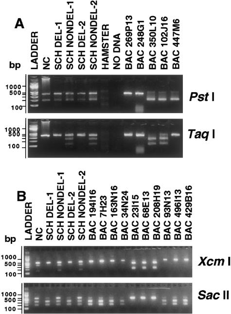
Williams-Beuren syndrome (WBS) is a developmental disorder caused by haploinsufficiency for genes in a 2-cM region of chromosome band 7q11.23. With the exception of vascular stenoses due to deletion of the elastin gene, the various features of WBS have not yet been attributed to specific genes. Although >/=16 genes have been identified within the WBS deletion, completion of a physical map of the region has been difficult because of the large duplicated regions flanking the deletion. We present a physical map of the WBS deletion and flanking regions, based on assembly of a bacterial artificial chromosome/P1-derived artificial chromosome contig, analysis of high-throughput genome-sequence data, and long-range restriction mapping of genomic and cloned DNA by pulsed-field gel electrophoresis. Our map encompasses 3 Mb, including 1.6 Mb within the deletion. Two large duplicons, flanking the deletion, of >/=320 kb contain unique sequence elements from the internal border regions of the deletion, such as sequences from GTF2I (telomeric) and FKBP6 (centromeric). A third copy of this duplicon exists in inverted orientation distal to the telomeric flanking one. These duplicons show stronger sequence conservation with regard to each other than to the presumptive ancestral loci within the common deletion region. Sequence elements originating from beyond 7q11.23 are also present in these duplicons. Although the duplicons are not present in mice, the order of the single-copy genes in the conserved syntenic region of mouse chromosome 5 is inverted relative to the human map. A model is presented for a mechanism of WBS-deletion formation, based on the orientation of duplicons' components relative to each other and to the ancestral elements within the deletion region.


Figures

Figure  1
Figure 1
STS-content map of BAC/PAC contig of WBS deletion and flanking regions. Red circles denote STS present by PCR; black circles denote STS absent by PCR; aqua circles denote STS present by sequence alignment with HTGS data; yellow circles denote STS absent by sequence alignment; white circles denote STS not assessed. Multicopy sequence clusters (REPs) are presented as colored bars above the contig. Arrows indicate orientation (arbitrarily defined) of these REPs relative to each other. The centromeric and telomeric breakpoint limits (black bars) were determined by SSN assays of hybrid cell lines derived from one individual with WBS.
Figure  2
Figure 2
Long-range restriction map of the WBS deletion and flanking repeat–containing regions, as derived from PFGE studies of BACs, human × hamster somatic-cell hybrids, and human lymphoblasts. The map covers 3 contiguous Mb, including 1.5–1.7 Mb within the deletion. The centromeric and telomeric breakpoint limits were determined by PFGE blot hybridization data. Restriction sites of methylation-sensitive enzymes identified in BACs but not in genomic DNA are indicated by asterisks (*). The few unexplained discrepancies between genomic and clone restriction sites are also indicated.
Figure  3
Figure 3
NotI fragment analysis of the flanking and deletion-junction regions. A, 2–4-Mb resolution Southern blot of the four WBS SCH lines, hybridized successively with flanking region probes. The probe for POM121, which is part of REP B, hybridized to both normal (3 and 1 Mb) NotI fragments and the 4-Mb junction fragment present only in the SCH DEL lanes. A probe for the telomeric locus SHGC-31781 hybridized to the lower fragment and to the junction fragment and thus localized the 1-Mb fragment to the telomeric side. The IB2070 probe from the centromeric flanking region recognized only the upper fragment and the junction fragment, consistent with the 3-Mb NotI fragment's being derived from the centromeric side. The HIP1 locus, previously localized outside the WBS deletion (Wedemeyer et al. 1997), was localized to the telomeric side, by hybridization to the 1-Mb fragment in the SCH NONDEL lanes and to the junction fragment in the SCH DEL lanes. B, Southern blot of seven human normal control samples (NC), an SCH DEL line, and an SCH NONDEL line, hybridized successively with probes for POM121 and GTF2I (IB291). Both probes recognized the 1- and 3-Mb bands flanking the deletion, as well as the 4-Mb deletion-junction fragment. For POM121, differential hybridization intensities suggest a 2:1 copy-number ratio for the 3-Mb fragment, relative to the 1-Mb fragment, whereas for GTF2I the relative gene dosage is reversed. C, Map of the NotI fragments flanking the WBS deletion and of the deletion-junction fragment. IB2070, SHGC-31781, and HIP1 are single-copy loci mapped to either the centromeric (IB2070) or the telomeric (SHGC-31781 and HIP1) flanking region. GTF2I and POM121 exist in three copies. Both probes hybridize to the 4-Mb fragment with greater intensity than do the single copy probes suggesting that two copies of these loci are flanking the deletion and that one is within the deletion. D, PFGE Southern blots containing samples from affected (WS) individuals, parental controls (PAT and MAT), and unrelated normal controls (NC), hybridized with either the POM121 or the GTF2I probes, documenting the consistency of the 4-Mb junction fragments in a larger number of individuals with WBS. As in B, both probes recognize the 1- and 3-Mb NotI fragments in all samples. The 4-Mb fragment is seen with either probe in all seven unrelated WS samples. The de novo occurrence of this deletion-junction fragment in case WS 1480 is illustrated by its absence in both parents.
Figure  4
Figure 4
A, Identification of the third GTF2I/NCF1 locus by restriction digestion of the 392-bp amplimer of NCF1 exon 3–4 from human cells, SCH, and BACs. These PCR products contain two SSN differences that change restriction-enzyme sites: a PstI site at position 213 of the 350L10 amplimer (A at 213) is absent on 269P13 (G at 213), and a TaqI site at position 262 of 350L10 (C at 263) is also absent on 269P13 (T at 263). Intact and digested PstI fragments were generated from both the deleted and nondeleted SCH lines. Amplimers of BACs 269P13 and 248G1 (GTF2IP1/NCF1P1) are not cut, whereas those of BACs 350L10 and 102J16 (GTF2I/NCF1) and 447M6 (GTF2IP2/NCF1P2) are. In contrast, TaqI-cleaved bands were apparent only in the SCH NONDEL-1 and -2 lanes, consistent with deletion of the NCF1 locus residing on BACs 350L10 and 102J16(GTF2I/NCF1), which also show TaqI digestion. Amplimers from BACs 269P13 and 248G1(GTF2IP1/NCF1P1) and BAC 447M6 (GTF2IP2/NCF1P2) are not cut, consistent with their mapping to loci outside the deletion. The combined TaqI and PstI data are consistent with a model placing BAC 447M6 at a third NCF1 locus, outside the deletion. Incomplete TaqI digestion of the BAC 102J16 amplimer does not affect the conclusions, because the presence of a single locus with the TaqI site has been confirmed by direct sequencing of the amplimer. B, Assignment of BACs to repeat clusters by restriction digestion of human, SCH, and BAC amplimers of the 479-bp STS 208H19M (in REP B1) containing two SSN differences. Sequence comparisons revealed the presence of an XcmI site at position 362 for the 208H19 amplimer (CT at 366–367) and its absence on that for 313P13 (2-bp deletion, relative to 208H19); in contrast, a SacII site was predicted at position 324 for 313P13 (T at 326) but not for 208H19 (G at 326). XcmI restriction digestion generated, for BACs 23I15, 68E13, and 208H19, predicted fragments of 330 and 150 bp that were absent in all other BACs tested. Bands of the same size were also generated from the SCH NONDEL lines but not from the SCH DEL lines. In contrast, SacII yielded predicted bands of 300 and 180 bp only for BACs 194I16, 7H23, 163N16, and 34N24. In the case of SacII, uncut fragments were of significantly lower intensity when they were from the SCH DEL lines. These results provide strong evidence for the presence of a locus on BACs 23I15, 68E13, and 208H19 within the WBS deletion, a locus that is highly homologous to extradeletion sequences on BACs 194I16, 7H23, 34N24, 93N13, 496I13, 429B16, and 163N16.

References

Electronic-Database Information

    1. GenBank, http://www.ncbi.nlm.nih.gov/Web/Genbank/Genbank/Overview.html
    1. Genome Sequencing Center, Washington University, St. Louis, http://genome.wustl.edu/gsc/
    1. Human Chromosome 7 Mapping and Sequencing, http://genome.nhgri.nih.gov/chr7/
    1. Online Mendelian Inheritance in Man (OMIM), http://www.ncbi.nlm.nih.gov/Omim/ (for WBS [MIM 194050], CGD [MIM 233700], VCFS [MIM 192430], SMS [MIM 182290], PWS [MIM 176270], AS [MIM 105830], HNPP [MIM 162500], SMAI, II, and III [MIM 253300, MIM 253550, and MIM 253400])
    1. Stanford Human Genome Center, http://www-shgc.stanford.edu/Mapping/index.html

References

    1. Altschul SF, Gish W, Miller W, Myers EW, Lipman DJ (1990) Basic local alignment search tool. J Mol Biol 215:403–410 - PubMed
    1. Altschul SF, Madden TL, Schaffer AA, Zhang J, Zhang Z, Miller W, Lipman DJ (1997) Gapped BLAST and PSI-BLAST: a new generation of protein database search programs. Nucleic Acids Res 25:3389–3402 - PMC - PubMed
    1. Amos-Landgraf JM, Ji Y, Gottlieb W, Depinet T, Wandstrat AE, Cassidy SB, Driscoll DJ, et al (1999) Chromosome breakage in the Prader-Willi and Angelman syndromes involves recombination between large, transcribed repeats at proximal and distal breakpoints. Am J Hum Genet 65:370–386 - PMC - PubMed
    1. Baumer A, Dutly F, Balmer D, Riegel M, Tukel T, Krajewska-Walasek M, Schinzel AA (1998) High level of unequal meiotic crossovers at the origin of the 22q11.2 and 7q11.23 deletions. Hum Mol Genet 7:887–894 - PubMed
    1. Bouffard GG, Idol JR, Braden VV, Iyer LM, Cunningham AF, Weintraub LA, Touchman JW, et al (1997) A physical map of human chromosome 7: an integrated YAC contig map with average STS spacing of 79 kb. Genome Res 7:673–692 - PubMed

Publication types

LinkOut - more resources