. 2000 Jan;66(1):128-35.
doi: 10.1086/302724.

Fibrodysplasia ossificans progressiva, a heritable disorder of severe heterotopic ossification, maps to human chromosome 4q27-31

Affiliations

Fibrodysplasia ossificans progressiva, a heritable disorder of severe heterotopic ossification, maps to human chromosome 4q27-31

G Feldman et al. Am J Hum Genet. 2000 Jan.

Abstract

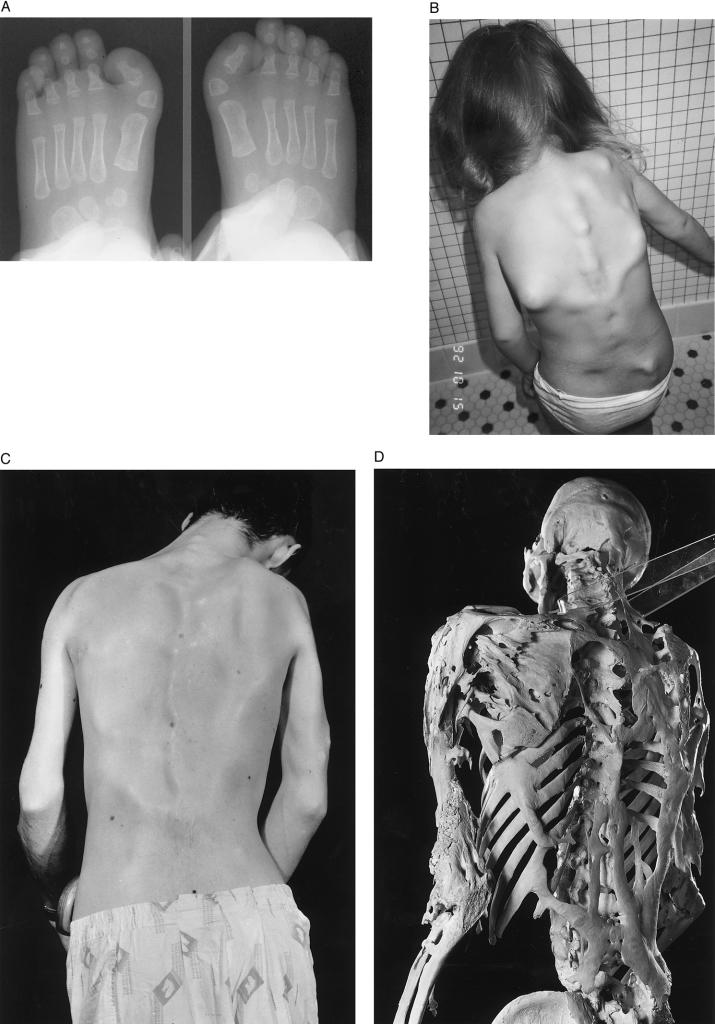
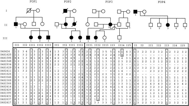
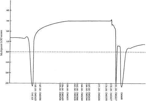
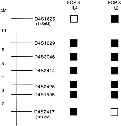
Fibrodysplasia ossificans progressiva (FOP) is a severely disabling, autosomal-dominant disorder of connective tissue and is characterized by postnatal progressive heterotopic ossification of muscle, tendon, ligament, and fascia and by congenital malformation of the great toes. To identify the chromosomal location of the FOP gene, we conducted a genomewide linkage analysis, using four affected families with a total of 14 informative meioses. Male-to-male transmission of the FOP phenotype excluded X-linked inheritance. Highly polymorphic microsatellite markers covering all human autosomes were amplified by use of PCR. The FOP phenotype is linked to markers located in the 4q27-31 region (LOD score 3.10 at recombination fraction 0). Crossover events localize the putative FOP gene within a 36-cM interval bordered proximally by D4S1625 and distally by D4S2417. This interval contains at least one gene involved in the bone morphogenetic protein-signaling pathway.


Figures

Figure 1
Figure 1
Clinical findings in FOP. A, Radiographs of the feet at age 4 mo in a child with FOP. The soft-tissue shadows clearly outline the short in-turned great toes due to malformed delta-shaped proximal phalanges (arrowheads), one of the common variations of great-toe malformations seen in FOP. B, Multiple variable-size soft-tissue nodules (preosseous fibroproliferative lesions) on the back of a 4-year-old child with FOP. These fibroproliferative lesions are frequently the first indication of heterotopic ossification in a child. C and D, Clinical appearance and skeleton of a man with FOP. The rigid posture in this 25-year-old man was due to ankylosis of the spine, shoulders, and elbows. He died of pneumonia at age 40 years. Plates and ribbons of ectopic bone contour the skin over the back and arms (C) and can be seen directly on the skeleton (D). (Courtesy of the Mutter Museum, College of Physicians of Philadelphia; reproduced with permission from Shafritz et al. [1996].)
Figure  2
Figure 2
Pedigrees of families with FOP with genotypes for markers from chromosome 4q27-31. Standard pedigree notation is used: circles and squares denote females and males, respectively; blackened symbols indicate affected individuals; and generations are indicated by Roman numerals to the left of the pedigrees (only individuals from whom genotyping data was obtained are numbered). Haplotypes for the markers shown on the left are listed below the symbols for the family members. The haplotype cosegregating with the disorder in each family is boxed.
Figure  3
Figure 3
Combined multipoint LOD scores for the markers in the 4q27-31 region in four families with FOP. Multipoint LOD scores were calculated for each of the markers indicated along the X-axis. The relative genetic distances of these markers, measured (in cM) from 4pter, were derived from the Research Genetics database.
Figure  4
Figure 4
Localization of the FOP-linked interval on the long arm of human chromosome 4. Schematic representation of the 4q27-31 region shows some of the markers used in the genetic linkage analysis. The genetic distances between the markers are indicated and the cumulative genetic distances (in cM; pter is 0 cM) for the markers defining the proximal and distal boundaries of the linked interval are shown in parentheses, under the marker names. Blackened squares denote regions of allele sharing among affected individuals; open squares represent recombinant regions. Crossovers in individual III-4 from family 3 identified the proximal boundary, and those in individual III-2 from family 3 identified the distal boundary.

References

Electronic-Database Information

    1. GeneMap 1999, http://www.ncbi.nlm.nih.gov/genemap/ (for localization of genes and expressed-sequence tags)
    1. Online Mendelian Inheritance in Man (OMIM), http://www.ncbi.nlm.nih.gov/Omim (for FOP [MIM 135100]) - PubMed
    1. Radiation Hybrid Mapping, http://carbon.mit.edu:8000/cgi-bin/contig/rhmapper.pl (for radiation-hybrid mapping)
    1. Research Genetics, Huntsville AL, http://www.resgen.com (for linkage analysis)

References

    1. Azimi N, Brown K, Bamford RN, Tagaya Y, Siebenlist U, Waldmann TA (1998) Human T cell lymphotropic virus type I Tax protein trans-activates interleukin 15 gene transcription through an NF-κB site. Proc Natl Acad Sci USA 95:2452–2457 - PMC - PubMed
    1. Bi W, Deng JM, Zhang Z, Behringer RR, De Crombrugghe B (1999) Sox9 is required for cartilage formation. Nat Genet 22:85–89 - PubMed
    1. Cohen RB, Hahn GV, Tabas JA, Peeper J, Levitz CL, Sando A, Sando N, et al (1993) The natural history of heterotopic ossification in patients who have fibrodysplasia ossificans progressiva. A study of forty-four patients. J Bone Joint Surg Am 75:215–219 - PubMed
    1. Connor JM, Evans DAP (1982a) Fibrodysplasia ossificans progressiva: the clinical features and natural history of 34 patients. J Bone Joint Surg Br 64:76–83 - PubMed
    1. ——— (1982b) Genetic aspects of fibrodysplasia ossificans progressiva. J Med Genet 19:35–39 - PMC - PubMed

Publication types

LinkOut - more resources