. 2000 Jan;122(1):35-48.
doi: 10.1104/pp.122.1.35.

Villin-like actin-binding proteins are expressed ubiquitously in Arabidopsis

Affiliations

Villin-like actin-binding proteins are expressed ubiquitously in Arabidopsis

U Klahre et al. Plant Physiol. 2000 Jan.

Abstract

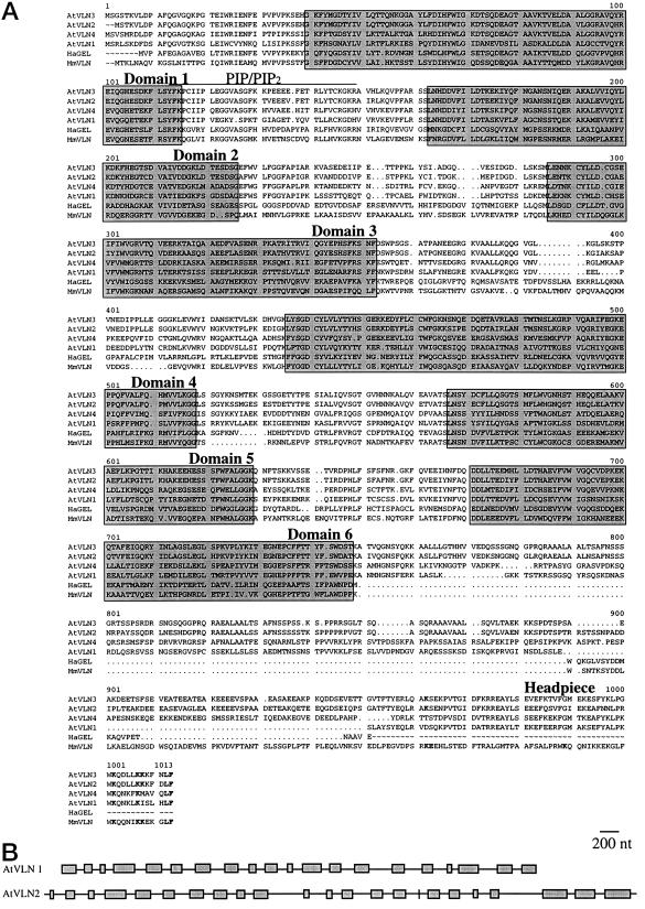
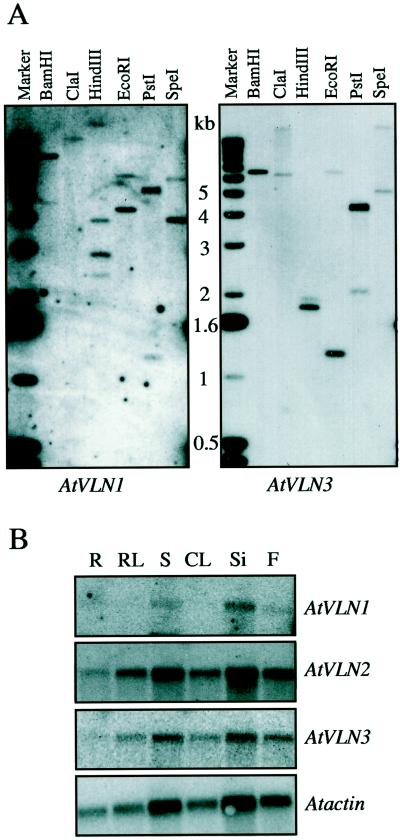
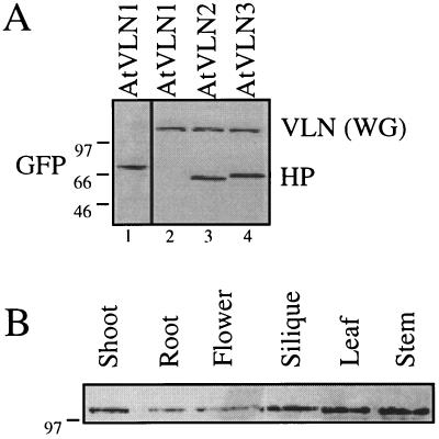
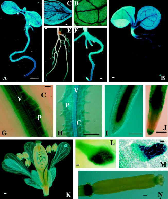
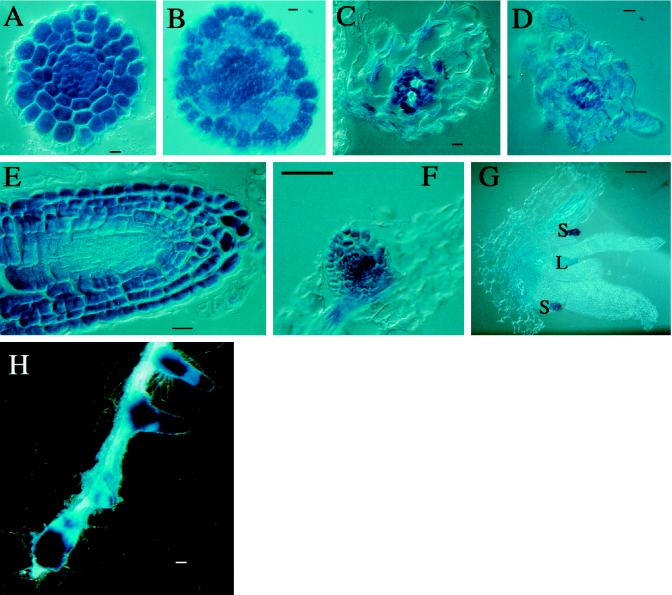
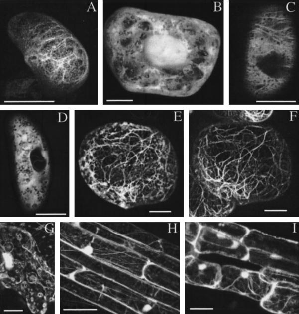
In an attempt to elucidate the biological function of villin-like actin-binding proteins in plants we have cloned several genes encoding Arabidopsis proteins with high homology to animal villin. We found that Arabidopsis contains at least four villin-like genes (AtVLNs) encoding four different VLN isoforms. Two AtVLN isoforms are more closely related to mammalian villin in their primary structure and are also antigenically related, whereas the other two contain significant changes in the C-terminal headpiece domain. RNA and promoter/beta-glucuronidase expression studies demonstrated that AtVLN genes are expressed in all organs, with elevated expression levels in certain types of cells. These results suggest that AtVLNs have less-specialized functions than mammalian villin, which is found only in the microvilli of brush border cells. Immunoblot experiments using a monoclonal antibody against pig villin showed that AtVLNs are widely distributed in a variety of plant tissues. Green fluorescent protein fused to full-length AtVLN and individual AtVLN headpiece domains can bind to both animal and plant actin filaments in vivo.


Figures

Figure 1
Figure 1
Deduced amino acid sequences and genomic structures of Arabidopsis VLN genes. A, Alignment of AtVLNs with animal villin and gelsolin. Four Arabidopsis amino acid sequences (AtVLN1–4) were compared with those of mouse villin (MmVLN) and lobster gelsolin (HaGEL). AtVLN 4 is a recent submission to GenBank (accession no. Y12782). The evolutionarily conserved domains 1 through 6 are boxed and indicated. Also indicated is the headpiece domain. Residues that are important for actin binding (Doering and Matsudaira, 1996) are printed in bold letters. The putative PIP/PIP2 binding domain is overlined. Note the major difference between animal villins and AtVLNs in the size of the linker domain between the core and the headpiece. This domain is rich in hydroxylated amino acids (18%–28%) and varies substantially between the isoforms. The sequences for cDNAs of AtVLNs 1 to 3 were deposited in GenBank and have the accession numbers. AF81201, AF81202, and AF81203, respectively. B, Genomic arrangement of AtVLN 1 and 2. Exons are shown as boxes and introns as lines.
Figure 2
Figure 2
Abundance and expression of AtVLN genes. A, Genomic DNA gel-blot hybridizations. Six micrograms of total Arabidopsis DNA digested with the indicated restriction enzymes were loaded per lane. The left blot was hybridized with a cDNA fragment of AtVLN 1 and the right blot with a cDNA fragment of AtVLN 3. B, RNA gel-blot hybridizations. One microgram of poly(A+) RNA was loaded per lane and the blot was hybridized with cDNA fragments as indicated. The blot was rehybridized with a genomic fragment of Arabidopsis ACTIN 1 as a loading control (AtACTI). The size of transcripts was approximately 3 kb for AtVLN and 1.6 kb for AtACT1.
Figure 3
Figure 3
Detection of AtVLN by a monoclonal antibody. A, A monoclonal antibody against pig villin headpiece (Dudouet et al., 1987) recognizes AtVLN 2 and 3, but not AtVLN 1. GFP-headpiece fusion proteins were synthesized by wheat germ in vitro translation reactions and treated on a western blot with an anti-villin antibody (HP; lanes 2–4). The presence of GFP-AtVLN 1 was verified using an antibody raised against GFP (GFP, lane 1, CLONTECH). The anti-villin antibody also recognizes VLN(WG) (the wheat germ VLN). B, AtVLNs can be detected in all organs. Western blots of crude protein extracts (approximately 30 μg per lane) were treated with the monoclonal anti-villin antibody. Molecular masses (in kD) are indicated on the left of the panels.
Figure 4
Figure 4
Expression pattern of representative AtVLN::GUS transgenic plants. A, C, E, G, I, K, L, and N are from AtVLN 1::GUS plants; B, D, F, H, J, and M are from AtLVN 2::GUS plants. A and B, Entire seedling. C and D, Cotyledon. E and F, Roots. G and H, Root, high magnification; C, cortex; P, pericycle; V, vasculature. I and J, Root tip, high magnification. K, Flower. L and M, Stipule. N, Silique. GUS staining was performed for 1 h in I, K, and N; for 2 h in E, I, F, J, and H; and for 24 h in B, D, and M. Scale bars represent 1 mm in A and E; 100 μm in B through D, F, H through K, and N; and 10 μm in G, L, and M.
Figure 5
Figure 5
Cell-specific expression in AtVLN 1::GUS (A, C, E, and G) and AtVLN 2::GUS (B, D, and F) transgenic plants. A and B, Cross-sections of root tips. C and D, Cross-sections through adult root. E, Longitudinal section of root tip. F, Longitudinal section of lateral root bud. G, Section through apical meristem; S, stipules; L, leaf primordium. GUS staining was performed for 16 h. H, Staining of a root of an AtVLN 1::GUS plant. Two days after treatment with 2,4-D, GUS staining was performed for 2 h. Scale bars represent 10 μm in A through E and 100 μm in F through H.
Figure 6
Figure 6
AtVLNs interact with F-actin in vivo. A, GFP-derived fluorescence from a BY2 cell expressing GFP-AtVLN 3. B, Fluorescence of a BY2 cell expressing GFP alone. C, Optical section through a cell expressing GFP-AtVLN 2-headpiece. D, Optical section through a cell expressing GFP alone. E, GFP-derived fluorescence from a BY2 cell expressing GFP-AtVLN 3-headpiece. F, Same cell as E, but counterstained with rhodamine-phalloidin. G through I, GFP-derived fluorescence from transgenic plant carrying GFP-AtVLN 1-headpiece. G, Epidermis; H, hypocotyl; I, root. Circular structures in epidermal cells were regularly observed, but are of unknown origin. Scale bar represents 10 μm in A through F and 50 μm in G through I.
Figure 7
Figure 7
Expression of GFP-fusion proteins in mammalian Vero cells. A, Rhodamine-phalloidin staining of B. B, GFP-headpiece-AtVLN3 fusion protein, dorsal face of cell, showing microvilli. C, Rhodamine-phalloidin staining of D. D, GFP-headpiece-AtVLN1 fusion protein, base of cell, showing membrane extensions. E, Rhodamine-phalloidin staining of F. F, GFP-headpiece-AtVLN3 fusion protein, base of cell, showing membrane extensions. G, Rhodamine-phalloidin staining of H. H, GFP-headpiece-AtVLN3 fusion protein, base of cell, showing stress fibers.

Similar articles

Cited by

References

    1. Ampe C, Vanderkerckhove J. The F-actin capping proteins of Physarum polycephalum: cap42(a) is very similar, if not identical, to fragmin and is structurally and functionally very homologous to gelsolin; cap42(b) is Physarum actin. EMBO J. 1987;6:4149–4157. - PMC - PubMed
    1. Andre E, Lottspeich F, Schleicher M, Noegel A. Severin, gelsolin, and villin share a homologous sequence in regions presumed to contain F-actin severing domains. J Biol Chem. 1988;263:722–727. - PubMed
    1. Aoyama T, Chua N-H. A glucocorticoid-mediated transcriptional induction system in transgenic plants. Plant J. 1997;11:605–612. - PubMed
    1. Arpin M, Pringault E, Finidori J, Garcia A, Jeltsch J-M, Vandekerckhove J, Louvard D. Sequence of human villin: a large duplicated domain homologous with other actin-severing proteins and a unique small carboxy-terminal domain related to villin specificity. J Cell Biol. 1988;107:1759–1766. - PMC - PubMed
    1. Barnes SA, Nishizawa NK, Quaggio RB, Whitelam GC, Chua N-H. Far-red light blocks greening of Arabidopsis seedlings via a phytochrome A-mediated change in plastid development. Plant Cell. 1996;8:601–615. - PMC - PubMed

Publication types