. 2000 Jan 15;20(2):558-67.
doi: 10.1523/JNEUROSCI.20-02-00558.2000.

Inflammatory mechanisms in Alzheimer's disease: inhibition of beta-amyloid-stimulated proinflammatory responses and neurotoxicity by PPARgamma agonists

Affiliations

Inflammatory mechanisms in Alzheimer's disease: inhibition of beta-amyloid-stimulated proinflammatory responses and neurotoxicity by PPARgamma agonists

C K Combs et al. J Neurosci. .

Abstract

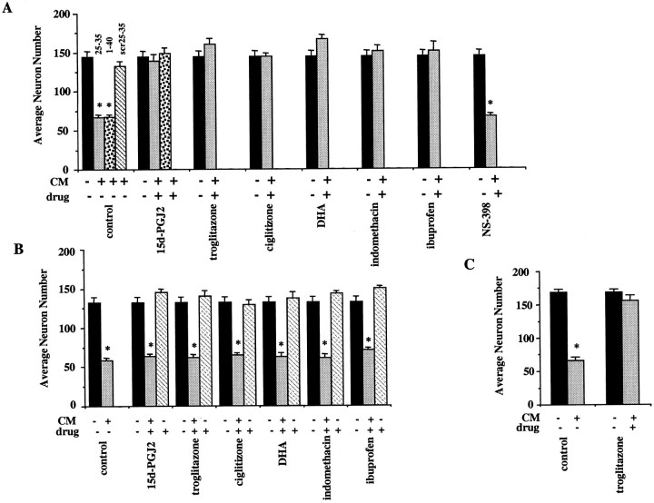
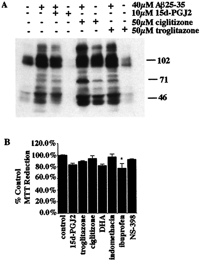
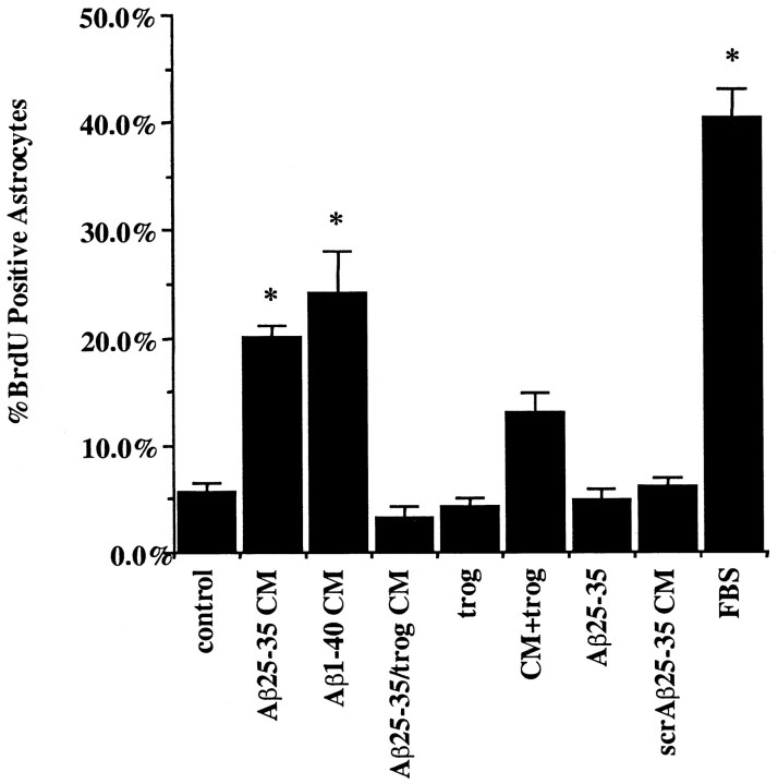
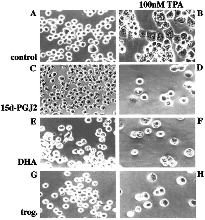
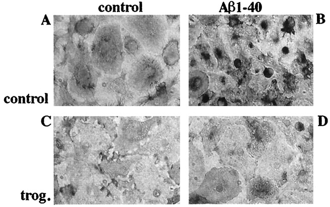
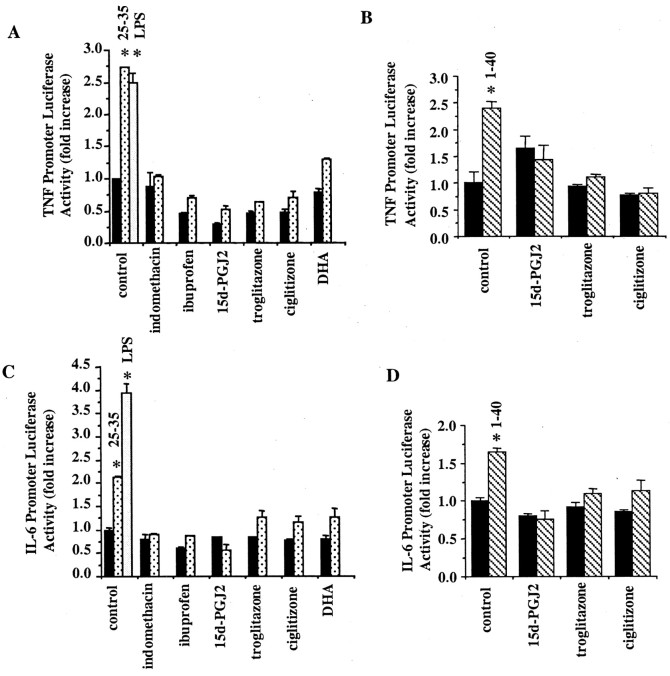
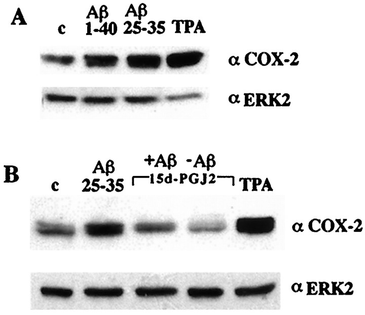
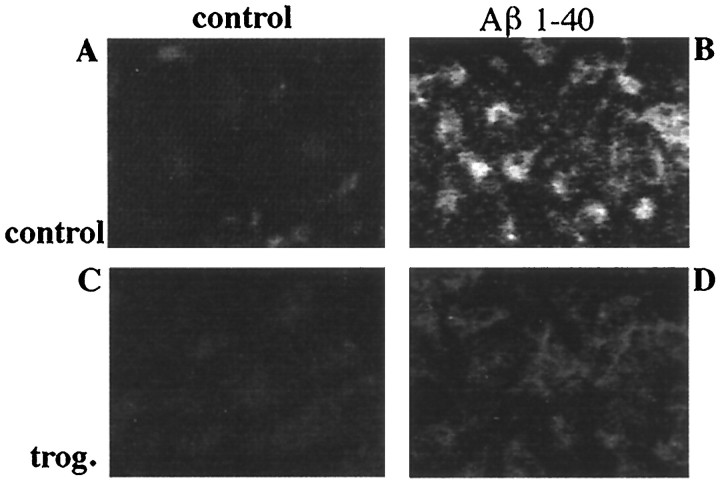
Alzheimer's disease (AD) is characterized by the extracellular deposition of beta-amyloid fibrils within the brain and the subsequent association and phenotypic activation of microglial cells associated with the amyloid plaque. The activated microglia mount a complex local proinflammatory response with the secretion of a diverse range of inflammatory products. Nonsteroidal anti-inflammatory drugs (NSAIDs) are efficacious in reducing the incidence and risk of AD and significantly delaying disease progression. A recently appreciated target of NSAIDs is the ligand-activated nuclear receptor peroxisome proliferator-activated receptor gamma (PPARgamma). PPARgamma is a DNA-binding transcription factor whose transcriptional regulatory actions are activated after agonist binding. We report that NSAIDs, drugs of the thiazolidinedione class, and the natural ligand prostaglandin J2 act as agonists for PPARgamma and inhibit the beta-amyloid-stimulated secretion of proinflammatory products by microglia and monocytes responsible for neurotoxicity and astrocyte activation. The activation of PPARgamma also arrested the differentiation of monocytes into activated macrophages. PPARgamma agonists were shown to inhibit the beta-amyloid-stimulated expression of the cytokine genes interleukin-6 and tumor necrosis factor alpha. Furthermore, PPARgamma agonists inhibited the expression of cyclooxygenase-2. These data provide direct evidence that PPARgamma plays a critical role in regulating the inflammatory responses of microglia and monocytes to beta-amyloid. We argue that the efficacy of NSAIDs in the treatment of AD may be a consequence of their actions on PPARgamma rather than on their canonical targets the cyclooxygenases. Importantly, the efficacy of these agents in inhibiting a broad range of inflammatory responses suggests PPARgamma agonists may provide a novel therapeutic approach to AD.


Figures

Fig. 1.
Fig. 1.
PPARγ agonists prevent neuronal death induced by β-amyloid-stimulated microglia and monocytes. THP-1 monocytes (A, B) or microglia (C) were stimulated for 48 hr with β-amyloid by plating the cells into uncoated wells (black bars) or wells coated with fibrillar Aβ25–35 (light gray bars) or Aβ1–40 (speckled bars) or nonfibrillar, scrambled Aβ25–35 (scr; negative control; striped bar) (all used at 48 pmol/mm2). A, C, The cells were cultured for 48 hr in the absence or presence of DMSO vehicle (control), 15d-PGJ2 (10 μm), troglitazone (50 μm), ciglitizone (50 μm), DHA (50 μm), indomethacin (100 μm), ibuprofen (600 μm), or NS-398 (5 μm). The conditioned media (CM) were collected, added to purified cultures of mouse cortical neurons (E16; 4.0 × 10⁴ neurons/well; 5 d in vitro), and incubated for 72 hr. The neuronal cultures were then fixed and stained for neuron-specific MAP2 protein, and surviving neurons were counted. B, In parallel experiments, the specificity of PPARγ ligand action for microglia and monocytes was demonstrated by plating THP-1 cells onto uncoated wells or wells coated with Aβ25–35 (light gray bars; 48 pmol/mm2) followed by incubation for 48 hr. The CM from the THP-1 cells stimulated with Aβ25–35 were collected and added to mouse cortical neurons (4.0 × 10⁴ neurons/well; 5 d in vitro) along with PPARγ ligands (light gray bars): 15d-PGJ2 (5 μm), troglitazone (25 μm), ciglitizone (25 μm), DHA (50 μm), indomethacin (100 μm), and ibuprofen (600 μm). Media from unstimulated THP-1 cells were added to neurons along with DMSO vehicle (black bars; control). PPARγ agonists alone were added to neurons as well (stripedbars). The neuronal cultures were stained for neuron-specific MAP2 protein, and surviving neurons were counted. The mean values shown (± SEM) are representative of four independent experiments. Unpaired ANOVA was performed with Tukey-Kramer post comparison to evaluate statistical significance (* =p < 0.001).
Fig. 2.
Fig. 2.
β-Amyloid fibrils activate a tyrosine kinase-dependent signaling cascade in microglial lineage cells that is unaffected by PPARγ agonists. A, The effect of PPARγ agonists on the activation of the Aβ-stimulated tyrosine kinase-signaling cascade was examined by incubating THP-1 cells for 24 hr with DMSO vehicle only or with 10 μm 15d-PGJ2, 50 μm ciglitizone, or 50 μmtroglitazone and then stimulating the cells with 60 μmfibrillar Aβ25–35 in suspension in serum-free media for 5 min. Cell lysates were examined by Western blot using the anti-phosphotyrosine antibody 4G10. B, The MTT reduction assay was performed on THP-1 cells stimulated with PPARγ agonists and NS-398 to evaluate the toxicity of these drugs. Cells were stimulated for 48 hr at 37°C with DMSO vehicle (control) or 10 μm 15d-PGJ2, 50 μm ciglitizone, 50 μm troglitazone, 50 μm DHA, 100 μm indomethacin, 600 μm ibuprofen, and 5 μm NS-398. MTT was added during the last 4 hr of stimulation. The percent control MTT reduction was calculated on the basis of the absorbance of the reduced MTT product formazan at 570 nm. Mean values are shown (± SEM) from an experiment performed in triplicate and independently repeated three times. Unpaired ANOVA was performed with Tukey-Kramer post comparison to evaluate statistical significance (* = p < 0.05).
Fig. 3.
Fig. 3.
PPARγ agonists prevent astrocyte proliferation induced by β-amyloid-stimulated monocytes. Conditioned medium was collected from THP-1 monocytes that had been incubated alone or were stimulated with fibrillar Aβ for 48 hr by plating onto fixed, surface-bound fibrillar Aβ25–35 or Aβ1–40 or nonfibrillar, scrambled Aβ25–35 (negative control) (48 pmol/mm2) in the presence of DMSO vehicle or troglitazone (50 μm). The conditioned medium (CM) was collected from the THP-1 cultures and applied to purified mouse astrocyte cultures for 72 hr. Treatments added to astrocyte cultures were as follows: conditioned medium from THP-1 cells only with DMSO vehicle (control), conditioned medium from Aβ25–35-stimulated THP-1 cells (Aβ25–35 CM), conditioned medium from Aβ1–40-stimulated THP-1 cells (Aβ1–40 CM), conditioned medium from THP-1 cells stimulated with Aβ25–35 in the presence of 50 μm troglitazone (Aβ25–35/trog CM), medium alone with 50 μm troglitazone (trog), conditioned medium from Aβ25–35-stimulated THP-1 cells added along with direct addition of 50 μmtroglitazone to astrocytes (CM+trog), medium alone with 20 μm Aβ25–35 (Aβ25–35), conditioned medium from scrambled Aβ25–35-stimulated THP-1 cells (scrAβ25–35 CM), and medium alone with 10% heat-inactivated FBS (FBS). Addition of FBS served as a positive control for mitogen-stimulated astrocyte cell division. The control for the effect of direct Aβ peptide stimulation of astrocytes was performed by adding, in suspension, 20 μm Aβ25–35 directly to astrocytic cultures for 72 hr. The controls for the effects of PPARγ agonists on the astrocytes themselves were performed by adding 50 μm troglitazone directly to astrocytes or adding 50 μm troglitazone directly to astrocyte cultures together with conditioned media from Aβ-stimulated THP-1 cells. The astrocytes were fixed and double-labeled for GFAP and BrdU. The numbers of BrdU-positive astrocytes were counted and graphed as a percentage of control. Shown are mean values (± SEM) from one of three representative experiments performed in duplicate. Unpaired ANOVA was performed with Tukey-Kramer post comparison to evaluate statistical significance (* = p < 0.001).
Fig. 4.
Fig. 4.
PPARγ agonists prevent differentiation of THP-1 monocytes into macrophages. THP-1 monocytes were incubated with vehicle only (control; DMSO and ethanol; A) or induced to differentiate into macrophages by treatment with 100 nm TPA for 48 hr (B, D, F, H) in the absence (A, B) or presence of 10 μm 15d-PGJ2 (C, D), 50 μmDHA (E, F), or 50 μm troglitazone (trog.; G, H).
Fig. 5.
Fig. 5.
Aβ-stimulated MAC-1 expression is inhibited by PPARγ agonists. Primary mouse microglia were plated onto uncoated culture wells (A, C) or surface-bound fibrillar Aβ1–40 (48 pmol/mm2; B, D) for 48 hr in the presence of vehicle (DMSO; A, B) or 10 μm troglitazone (trog.; C, D). Cells were fixed and stained for MAC-1. Immunoreactivity was visualized using 3,3′-diaminobenzidine tetrahydrochloride as the chromogen.
Fig. 6.
Fig. 6.
PPARγ agonists inhibit IL-6 and TNFα gene expression. THP-1 cells were transiently transfected with TNFα–luciferase reporter (A, B) or IL-6–luciferase reporter (C, D) constructs and assayed for promoter activity 48 hr later. The cells were cotransfected with a β-galactosidase–reporter construct to control for transfection efficiency. During the last 4 hr the cells were incubated alone (blackbars), with fibrillar Aβ1–40 (40 μm;stripedbars;B, D) or Aβ25–35 (60 μm;speckledbars; A, C), or with 5 μg/ml LPS (positive control; open bars) in the presence or absence of troglitazone (20 μm), ciglitizone (50 uM), DHA (100 μm), 15d-PGJ2 (50 μm), ibuprofen (3 mm), or indomethacin (200 μm). The data shown represent the average (± SEM) of three independent experiments. Unpaired ANOVA was performed with Tukey-Kramer post comparison to evaluate statistical significance (* =p < 0.001).
Fig. 7.
Fig. 7.
Aβ-stimulated COX-2 expression in THP-1 cells is inhibited by PPARγ agonists. A, THP-1 monocytes were incubated alone (c), with fibrillar Aβ25–35 (60 μm) or Aβ1–40 (60 μm) in suspension, or with 100 nm TPA for 18 hr in serum-free RPMI, and COX-2 expression was monitored by Western analysis of cellular lysates using an anti-COX-2-specific antibody. The blots were reprobed using an anti-ERK2 antibody as a control for protein loading. B,THP-1 cells were incubated in vehicle alone (c; DMSO) or stimulated with fibrillar Aβ25–35 (60 μm) or TPA (100 nm) in the presence or absence of the PPARγ agonist 15d-PGJ2 (50 μm).
Fig. 8.
Fig. 8.
Aβ-stimulated COX-2 expression in primary microglia is inhibited by PPARγ agonists. Primary mouse microglia were plated onto underivatized culture wells (A, C) or surface-bound Aβ1–40 (48 pmol/mm2; B, D) for 48 hr in the presence of vehicle (DMSO; A, B) or 10 μm troglitazone (trog.;C, D). Cells were fixed and stained for COX-2. Immunoreactivity was visualized using Cy3-conjugated rabbit anti-mouse antibody.

Similar articles

Cited by

References

    1. Aisen PS. Inflammation and Alzheimer's disease: mechanisms and therapeutic strategies. Gerontology. 1997;43:143–149. - PubMed
    1. Banati RB, Gehrmann J, Schubert P, Kreutzberg GW. Cytotoxicity of microglia. Glia. 1993;7:111–118. - PubMed
    1. Berg L, McKeel DW, Jr, Miller JP, Baty J, Morris JC. Neuropathological indexes of Alzheimer's disease in demented and nondemented persons aged 80 years and older. Arch Neurol. 1993;50:349–358. - PubMed
    1. Bjorkman DJ. The effect of aspirin and nonsteroidal anti-inflammatory drugs on prostaglandins. Am J Med. 1998;105:8S–12S. - PubMed
    1. Bolten WW. Scientific rationale for specific inhibition of COX-2. J Rheumatol Suppl. 1998;51:2–7. - PubMed

Publication types

MeSH terms

Substances