. 2000 Jan 15;20(2):674-84.
doi: 10.1523/JNEUROSCI.20-02-00674.2000.

Nerve injury induces gap junctional coupling among axotomized adult motor neurons

Affiliations

Nerve injury induces gap junctional coupling among axotomized adult motor neurons

Q Chang et al. J Neurosci. .

Abstract

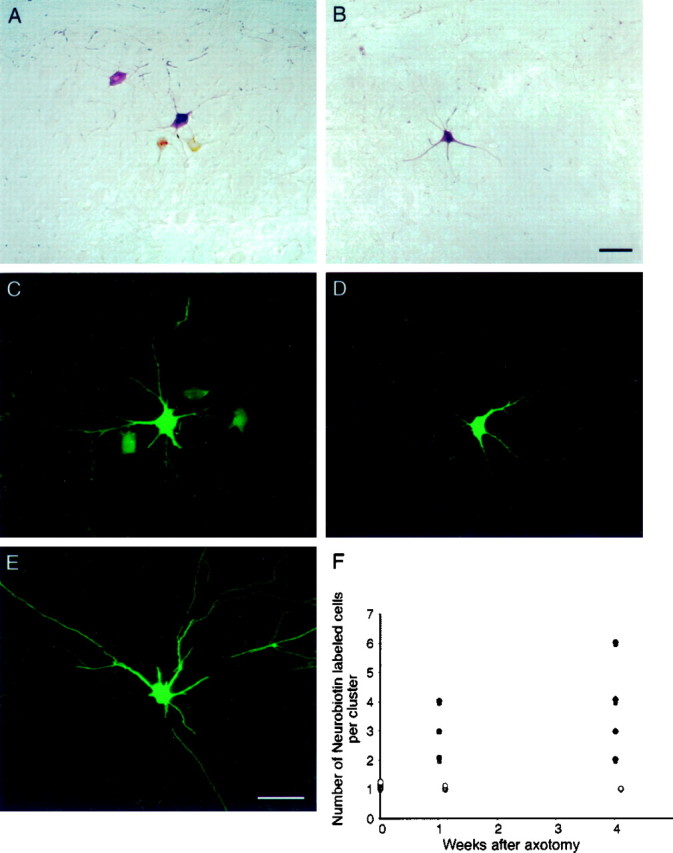
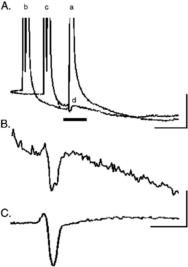
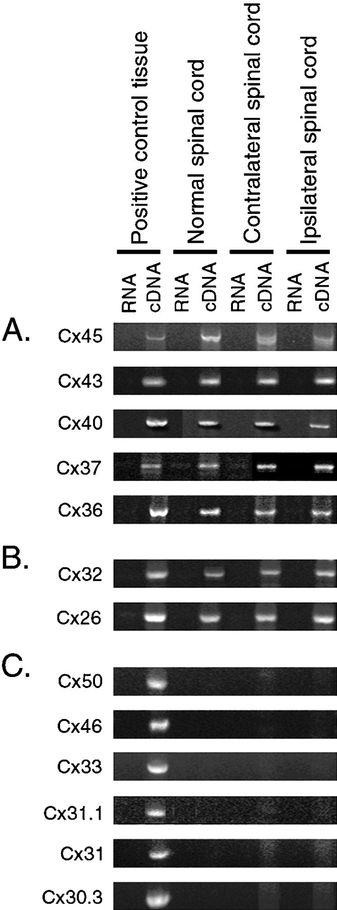
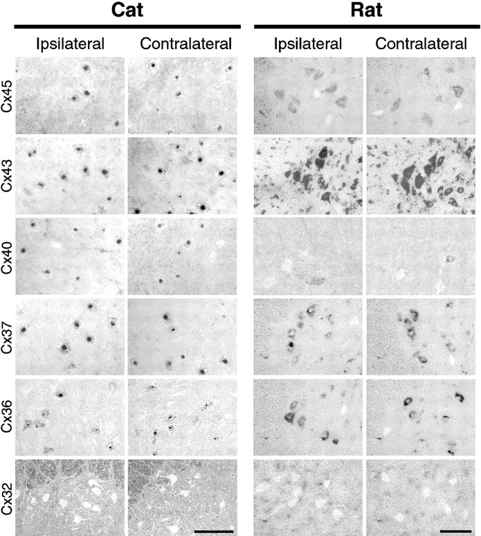
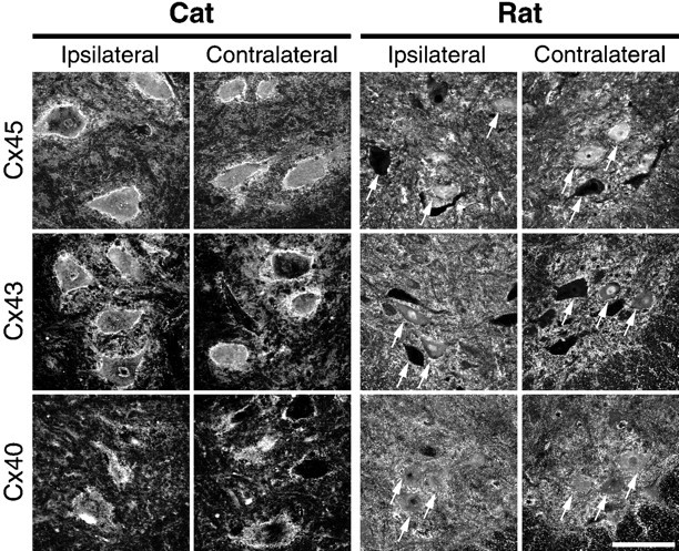
Neonatal spinal motor neurons are electrically and dye-coupled by gap junctions, but coupling is transient and disappears rapidly after birth. Here we report that adult motor neurons become recoupled by gap junctions after peripheral nerve injury. One and 4-6 weeks after nerve cut, clusters of dye-coupled motor neurons were observed among axotomized, but not control, lumbar spinal motor neurons in adult cats. Electrical coupling was not apparent, probably because of the electrotonic distance between dendrodendritic gap junctions and the somatic recording location. Analyses of gap junction protein expression in cat and rat showed that the repertoire of connexins expressed by normal adult motor neurons, Cx36, Cx37, Cx40, Cx43, and Cx45, was unchanged after axotomy. Our results suggest that the reestablishment of gap junctional coupling among axotomized adult motor neurons may occur by modulation of existing gap junction proteins that are constitutively expressed by motor neurons. After injury, interneuronal gap junctional coupling may mediate signaling that maintains the viability of axotomized motor neurons until synaptic connections are reestablished within their targets.


Figures

Fig. 1.
Fig. 1.
Axotomized motor neurons are extensively dye-coupled. A, Single plane projection of confocal stack of images showing a cluster of Neurobiotin-labeled motor neurons after injection of a single medial gastrocnemius motor neuron in a cat axotomized 1 week previously. Neurobiotin was revealed with HRP-conjugated streptavidin and a chromogenic reaction for HRP. There were a total of four labeled cells in this cluster, which occupied a region 214 × 234 × 189 μm in the dorsoventral, mediolateral, and rostrocaudal dimensions, respectively.B, Single plane projection of confocal stack of images showing a single cell body and a portion of the dendritic arbor of an injected peroneous motor neuron from a cat axotomized 1 week previously. Neurobiotin was revealed with fluorescein-conjugated streptavidin (also C–E). The absence of dye coupling in nonaxotomized motor neurons in animals in which the gastrocnemius–soleus nerve had been severed argues that gap junctional coupling is only observed among injured neurons. Scale bar:A, B, 100 μm. C, Single plane projection of confocal stack of images showing a cluster of Neurobiotin-labeled motor neurons after injection of a single medial gastrocnemius motor neuron in a cat axotomized 4 weeks previously. There were a total of four labeled cells in this cluster, which occupied a region 260 × 220 × 125 μm in the dorsoventral, mediolateral, and rostrocaudal dimensions, respectively.D, Single plane projection of confocal stack of images showing a single cell body and a portion of the dendritic arbor of an injected, nonaxotomized peroneous motor neuron from a cat axotomized 4 weeks previously. The absence of dye coupling in nonaxotomized motor neurons in animals in which the gastrocnemius–soleus nerve had been severed argues that gap junctional coupling is only observed among injured neurons, even at long times after axotomy. Scale bar:C–E, 100 μm. E, Single plane projection of confocal stack of images showing a single cell body and a portion of the dendritic arbor of an injected medial gastrocnemius motor neuron from a normal adult cat, showing the absence of dye coupling. F, Summary of the number of Neurobiotin-labeled cells per cluster in normal spinal cord in spinal cord 1 and 4–6 weeks after axotomy (filled circles) and in nonaxotomized motor pools in spinal cord 1 and 4–6 weeks after axotomy (open circles).
Fig. 2.
Fig. 2.
Electrical coupling among axotomized motor neurons is undetectable. Motor neurons were identified by intracellular impalement in cat spinal cord by the presence of an antidromic action potential (A, a) after muscle nerve stimulation. A, Intracellular current injection was used to elicit an orthodromic action potential (b) in the impaled motor neuron. By decreasing the interval between antidromic and orthodromic stimulation (c), the antidromic action potential elicited by ventral root stimulation failed (d). This failure occurs by collision of the antidromic spike by the intracellularly evoked action potential, which transiently inactivates voltage-gated sodium channels. Calibration: 10 mV, 5 msec. B, The region indicated by theblack line in the intracellular recording inA is shown at higher gain during collision. A depolarizing potential occurring within 5–10 msec of the antidromic stimulus artifact would be a putative coupling potential, but such potentials were not consistently observed. C, Extracellularly recorded field potential after antidromic ventral root stimulation (average of 5 sweeps) was subtracted from the intracellular trace during collision (B) to determine whether any remaining depolarizing potential was present. Calibration:B, C, 2 mV, 2 msec.
Fig. 3.
Fig. 3.
RT-PCR analysis of connexins expressed by normal adult and axotomized motor neurons. Using primers for each of the 13 known rodent connexins, PCR analysis was performed on cDNA from rat positive control tissues, such as heart (for Cx37, Cx40, Cx43, and Cx45), liver (for Cx26 and Cx32), eye (for Cx36, Cx46, and Cx50), skin (for Cx31.1, Cx30.3, and Cx31), and testis (for Cx33), normal spinal cord and spinal cord ipsilateral and contralateral to sciatic nerve cut 2 weeks previously. Corresponding RNAs were used as a negative controls. In each case, PCR products were eluted from gels, cloned, and sequenced to verify their identity. RNA was not pooled across animals, and the results shown here from one animal are representative of the other animals evaluated. A, B, Primers specific for Cx26, Cx32, and Cx36 amplified 364, 385, and 979 bp bands, respectively, from normal spinal cord, spinal cord contralateral and ipsilateral to sciatic nerve cut, and positive control tissue cDNA (Condorelli et al., 1998). Primers specific for Cx37, Cx40 (Haefliger et al., 1992), Cx43 (Beyer et al., 1987), and Cx45 (Schwarz et al., 1992) amplified 422, 308, 292, and 1217 bp bands, respectively, from normal spinal cord and spinal cord contralateral and ipsilateral to sciatic nerve cut and positive control tissue cDNA. Although Cx32 and Cx26 are detected by RT-PCR from spinal cord cDNA (B), in situ hybridization showed that these are not expressed by motor neurons (see Fig. 4).C, In contrast, primers against the other known rodent connexins amplified the predicted size band from positive control tissue but failed to amplify the same sized band in spinal cord. These results suggest that the repertoire of connexins expressed in lumbar spinal cord is unchanged after axotomy.
Fig. 4.
Fig. 4.
Connexin expression in motor neurons analyzed byin situ hybridization in rat and cat spinal cord is unchanged after axotomy. To compare the pattern of connexin expression between normal and axotomized rat motor neurons with normal and axotomized cat motor neurons in which extensive dye coupling had been characterized, in situ hybridization was performed with rat connexin cRNA probes and was visualized with a chromogenic reaction that resulted in positive cells appearing dark. Left, Shown are photographs of cat ventral L6–L7 spinal cord containing the gastrocnemius–soleus motor pools ipsilateral (left) or contralateral (left middle) to muscle nerve cut 4 weeks previously. Scale bar, 500 μm. Right, Shown are photographs of the lateral region of rat ventral spinal cord containing the sciatic nerve motor pools ipsilateral (right middle) or contralateral (right) to sciatic nerve cut 1 week previously. Cx45, Cx43, Cx40, Cx37, and Cx36 mRNA were detected in cat and rat motor neurons, identified by their location and large soma size. The pattern observed with each connexin probe after in situ hybridization in cat spinal cord was similar to that observed in rat for each experimental condition, with the exception that Cx40 was expressed in ∼10% of rat motor neurons compared with ∼75% of cat motor neurons. Neither Cx32 (bottom) or Cx26 (data not shown) were detected in motor neurons, but these were detected in meningeal and ependymal cells and glia. Scale bar, 100 μm.
Fig. 5.
Fig. 5.
Connexin protein expression in motor neurons is unchanged after axotomy in rat and cat spinal cord. To determine whether connexin proteins are expressed in rat and cat motor neurons, immunostaining with antibodies specific for Cx45, Cx43, and Cx40 was performed in cat (axotomized side, left; contralateral side, left middle) and rat (axotomized side, right middle; contralateral side,right) spinal cord. Shown are single plane projections of confocal stacks of images from cat L6–L7 dorsolateral ventral spinal cord containing the gastrocnemius motor pools or rat L3–L6 lateral and ventral spinal cord containing the sciatic nerve motor pools, obtained on a Leica TCS-4D system with a 40×, 1.25 NA oil immersion lens. In both rat and cat, anti-Cx45 (top row), Cx43 (middle row), and Cx45 (bottom row) antibodies revealed punctate staining surrounding motor neuron soma (several are indicated with arrows in each rat panel) and primary dendrites in the ventral horn. Some motor neurons had prominent cytoplasmic staining in addition to punctate, membrane-associated staining. Similar patterns of staining were observed in unmanipulated control animals (data not shown) and were similar in the spinal cord ipsilateral and contralateral to sciatic nerve cut 1 and 4–6 weeks after axotomy. In the middle row, note the absence of a change in Cx43 protein expression in glia in and around axotomized motor neurons (but see Rohlmann et al., 1993, 1994). These results show that, in rat and in cat, axotomy does not result in a detectable change in connexin protein expression in injured motor neurons. Scale bar, 100 μm.

Similar articles

Cited by

References

    1. Aldskogius H, Kozlova EN. Central neuron–glial and glial–glial interactions following axon injury. Prog Neurobiol. 1998;55:1–26. - PubMed
    1. Baker R, Llinas R. Electrotonic coupling between neurones in the rat mesencephalic nucleus. J Physiol (Lond) 1971;212:45–63. - PMC - PubMed
    1. Balice-Gordon RJ, Pereda A, Pinter MJ. Functional gap junctions couple motor neurons in development and reinnervation. Soc Neurosci Abstr. 1996;22:1487.
    1. Barry JA, Ribchester RR. Persistent polyneuronal innervation in partially denervated rat muscle after reinnervation and recovery from prolonged nerve conduction block. J Neurosci. 1995;15:6327–6339. - PMC - PubMed
    1. Beyer EC, Paul DL, Goodenough DA. Connexin43: a protein from rat heart homologous to a gap junction protein from liver. J Cell Biol. 1987;105:2621–2629. - PMC - PubMed

Publication types

MeSH terms

LinkOut - more resources