. 2000 Jan 15;20(2):685-95.
doi: 10.1523/JNEUROSCI.20-02-00685.2000.

Hebbian mechanisms revealed by electrical stimulation at developing rat neuromuscular junctions

Affiliations

Hebbian mechanisms revealed by electrical stimulation at developing rat neuromuscular junctions

G Busetto et al. J Neurosci. .

Abstract

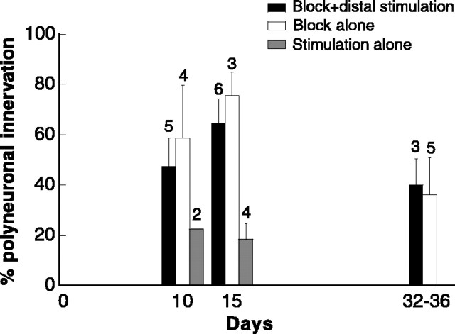
Synapse competition and elimination are widespread developmental processes, first demonstrated at neonatal neuromuscular junctions. Action potential activity was long shown to exert a powerful influence, but mechanisms and contribution relative to other factors are still not well understood. Here we show that replacement of natural motoneuronal discharge with synchronous activity suppresses elimination of polyneuronal innervation of myofibers. This requires the simultaneous chronic conduction block (tetrodotoxin) and distal electrical stimulation of motor axons during ectopic synaptogenesis in denervated adult soleus muscle. If in fact chronic stimulation is applied without central block of motor axons, the time course of synapse elimination is as fast as in control muscles undergoing natural activity. Our findings follow the prediction of Hebb's postulate and imply that asynchronous activity drives developmental synapse elimination in muscle. They further suggest that motoneurons could become transiently synchronized during development and regeneration, helping to establish the initial polyneuronal innervation.


Figures

Fig. 1.
Fig. 1.
Example of a polyneuronally innervated fiber recorded in the region of the fibular nerve growth, showing three steps of the EPP evoked by graded stimulation of fibular nerve under curare (a, superimposed traces) and the components of the full-sized potential of the same fiber obtained, in isolation, by computer subtraction (b) (see Materials and Methods for details).
Fig. 2.
Fig. 2.
Synchronous activity, evoked by electrical stimulation, inhibits synapse elimination in newly formed ectopic neuromuscular junctions in adult rat soleus muscle.a, Electrophysiological comparison of the amounts of polyneuronal innervation of myofibers is made at the indicated times after bilateral section of the original nerve, which activates synaptogenesis by the transplanted fibular nerve. Experimental side (filled circles): TTX conduction block + distal stimulation of fibular axons. Control contralateral side (open circles): fibular axons undergoing natural impulse activity. Each data point represents the percentage (mean ± SEM over several muscles) of polyneuronally innervated fibers (i.e. fibers with two or more steps of the evoked EPP, see Materials and Methods).Numbers above symbols indicate muscles. Five to thirty fibers per muscle (mean, 16.3; valid also for Fig. 3). *p < 0.05, **p < 0.005,t test. Exceptions to the above specifications: 19 of 34 control muscles are contralateral not to experimental muscles but to sides with block or stimulation alone or are unilateral preparations; they also had low values of polyneuronal innervation and were thus pooled together with the others. b, Comparison of the effects induced in different experimental muscles by changing the total number of stimuli per day shows that all amounts tested are effective; average time after section of soleus axons was comparable for the various amounts (19.3, 14.4, 18.6 d for 11,520, 34,560, and 86,400, respectively). Furthermore, the increase in synapse elimination is apparently equivalent regardless of the frequency of the stimuli in the train. The dashed line indicates mean level of polyneuronal innervation in the control muscles shown ina.
Fig. 3.
Fig. 3.
Comparison of the experimental muscles with similar muscles in which either stimulation or block has been omitted. Note that the muscles that were blocked but not stimulated had high levels of polyneuronal innervation. Stimulation alone, in contrast, did not prevent the elimination process. Pooling up the data at 10 and 15 d, the difference between experimental and stimulated alone muscles is highly statistically significant (p < 0.001). Other indications as in Figure2. Note that stimulated preparations without block had a sham cuff perfusing the sciatic nerve with saline, to control for nerve damage by the blocking technique: partial denervation of fibular nerve-reinnervated myofibers would complicate the interpretation of our findings, especially in blocked muscles, because the muscle effects of partial denervation (Cangiano and Lutzemberger, 1980) could induce sprouting and enhance polyneuronal innervation. Against its occurrence is: (1) nonblocked stimulated muscles with sham cuffs actually exhibit a low percentage of polyneuronal innervation, (2) fibular-evoked twitches in preparations with cuffs are not smaller than in those without cuffs (see Materials and Methods), and (3) extensive investigations of nerves with cuffs identical to those used here, indicated lack of damage (Pasino et al., 1996).
Fig. 4.
Fig. 4.
Cross sections of experimental (b), control (c), and purely paralyzed (d) soleus muscles, all reinnervated by the transplanted fibular nerve, showing that in the first two conditions large fibers are present on the side of the foreign nerve outgrowth, whereas in the latter only atrophic fibers are visible. a, Placement of cross sections:1, region of fibular nerve outgrowth, used for the experimental muscles in b and 2, center of muscle belly, used for muscles in c andd and to collect the data shown in Figure 5. Inb, muscle fibers (red) and foreign axons (green) are visualized with fluorescence microscopy (Materials and Methods). Stimulation parameters: 15 d, 87.5 msec trains, 7.5 trains per minute, train pulse frequency, 80 Hz, 86,400 pulses per day; 22 d, 467 msec, 7.5 trains per minute, 15 Hz, 86,400 pulses per day; 32 d, 87.5 msec, 1 train per minute, 80 Hz, 11,520 pulses per day. Two populations of myofibers are visible: (1) large fibers occupying most of the muscle cross-sectional area, in the region under nerve implant and of axonal penetration (small green spots); (2) atrophic fibers far from the nerve implant, especially tiny at margins of the muscle at 32 d (arrows), interpreted as not yet reinnervated fibers. Atrophic fibers are usually present on the entire side opposite to that of nerve penetration, but most of this side is not visible in the muscle at 32 d because it has been removed before the electrophysiological experiment, to speed up penetration of curare.c, d, Stain for myofibers as above, except biotin was revealed with peroxidase-avidin. *Side of implantation of fibular nerve. Scale bar, 500 μm.
Fig. 5.
Fig. 5.
Comparison of frequency distribution of fiber cross-sectional areas indicates complete similarity between experimental (block + distal stimulation) and control muscles (i.e. undergoing natural activity). Only blocked, only stimulated, and normal preparations are also shown, for comparison.a, All fibers present in the entire transverse section through the muscle center (Fig. 4a2) of three muscles for each condition, except Normal in which only some groups of fibers are sampled from one normal soleus. b, Only first layer fibers of the same sections, again from three muscles for each condition (Normal, one muscle), on the side of fibular nerve outgrowth. Arrow indicates a value = mean + 2 SDs for purely paralyzed muscles which is used to separate the population of large fibers in experimental and control muscles. All treatments lasting 32–34 d. Muscles of the conditions block + stimulation, control, and block alone were also used for electrophysiology, data of Figures 2 and 3; all muscles with systemic TTX, except stimulated alone ones.
Fig. 6.
Fig. 6.
Morphological and functional data (EPPs) of soleus fibers undergoing multiple innervation by the transplanted fibular nerve. a, Longitudinal muscle sections illustrate fibers with multiple synaptic sites of experimental andcontrol muscles at 15 d after section of soleus axons, as well as fibers not yet reinnervated of an experimental muscle at the same time delay. Axons and terminals are stained dark brown, and ACh-esterase are stained blue.b, Maximum apparent diameter measured in populations of fibers from experimental muscles (80 fibers with multiple synaptic sites), control muscles (83 fibers with single and multiple sites), and from denervatedfibers of both muscle types (80 fibers, devoid of NMJs and near muscle surface opposite to fibular nerve outgrowth). c, Class distribution of EPP durations (rise time + half relaxation time; see Materials and Methods and Fig. 1b) at 15 and 32–36 d, in polyneuronally innervated fibers of five experimentalmuscles (78 components, 38 fibers, 6.43 ± 0.27 msec, mean ± SEM), 11 control muscles (63 components, 30 fibers, 6.54 ± 0.33 msec) and five purely blocked muscles (73 components, 32 fibers, 8.24 ± 0.32 msec, significant difference with respect to both experimental and control muscles;p < 0.0005). Scale bar, 30 μm.

References

    1. Balice-Gordon RJ, Lichtman JW. Long-term synapse loss induced by focal blockade of postsynaptic receptors. Nature. 1994;372:519–524. - PubMed
    1. Barry JA, Ribchester RR. Persistent polyneuronal innervation in partially denervated rat muscle after reinnervation and recovery from prolonged nerve conduction block. J Neurosci. 1995;15:6327–6339. - PMC - PubMed
    1. Bennett MR, Pettigrew AG. The formation of synapses in striated muscle during development. J Physiol (Lond) 1974;241:515–545. - PMC - PubMed
    1. Benoit P, Changeux JP. Consequences of tenotomy on the evolution of multiinnervation in developing rat soleus muscle. Brain Res. 1975;99:354–358. - PubMed
    1. Betz WJ, Caldwell JH, Ribchester RR. The effects of partial denervation at birth on the development of muscle fibres and motor units in rat lumbrical muscle. J Physiol (Lond) 1980;303:265–279. - PMC - PubMed

Publication types

Grants and funding

LinkOut - more resources