. 2000 Jan 15;20(2):709-21.
doi: 10.1523/JNEUROSCI.20-02-00709.2000.

A highly conserved enhancer in the Dlx5/Dlx6 intergenic region is the site of cross-regulatory interactions between Dlx genes in the embryonic forebrain

Affiliations

A highly conserved enhancer in the Dlx5/Dlx6 intergenic region is the site of cross-regulatory interactions between Dlx genes in the embryonic forebrain

T Zerucha et al. J Neurosci. .

Abstract

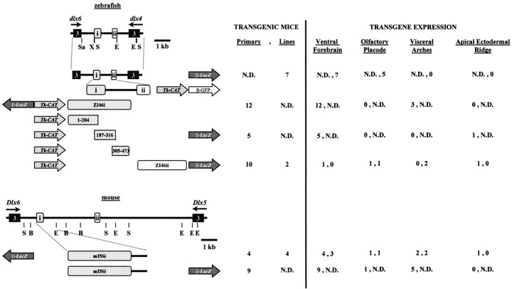
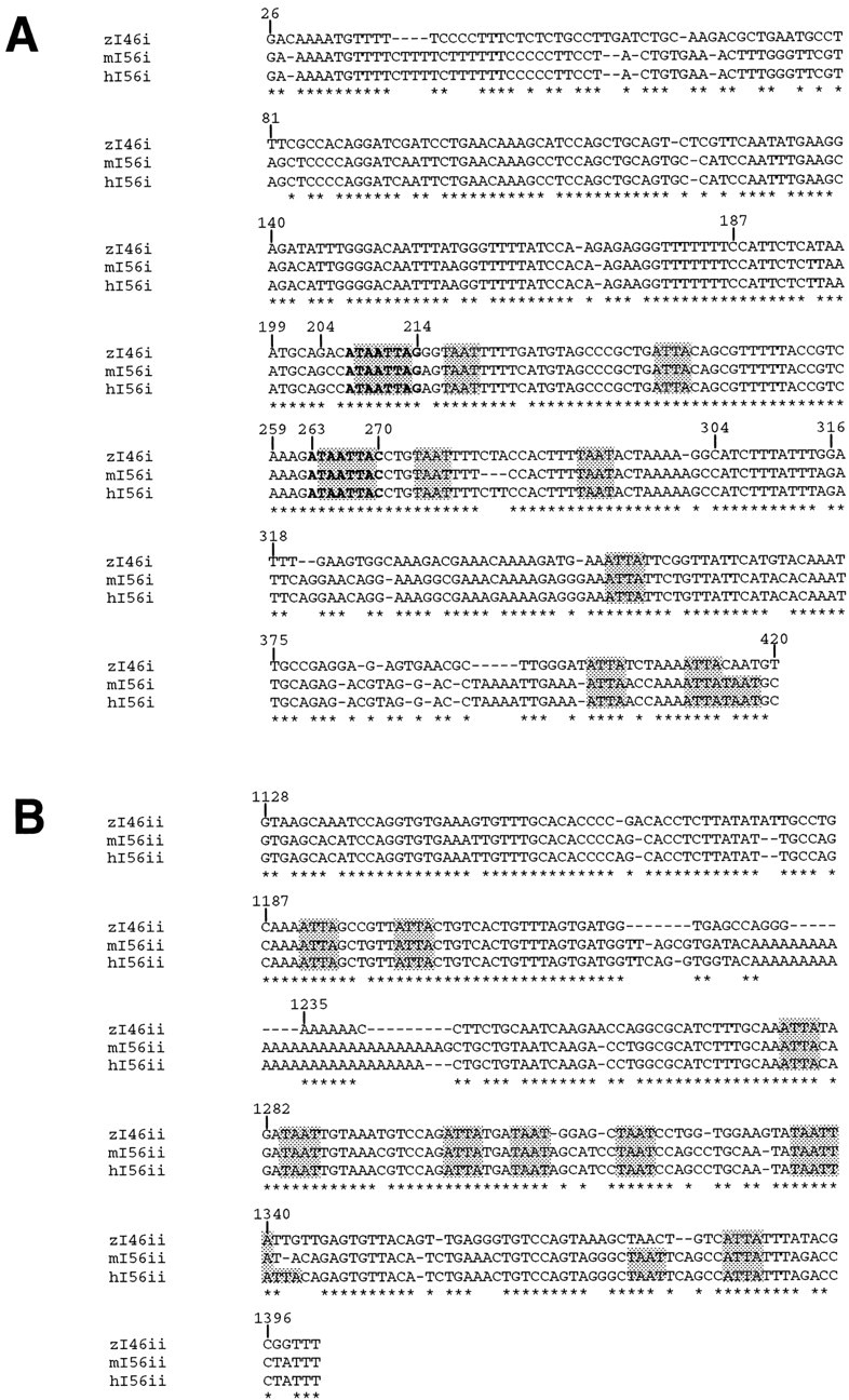
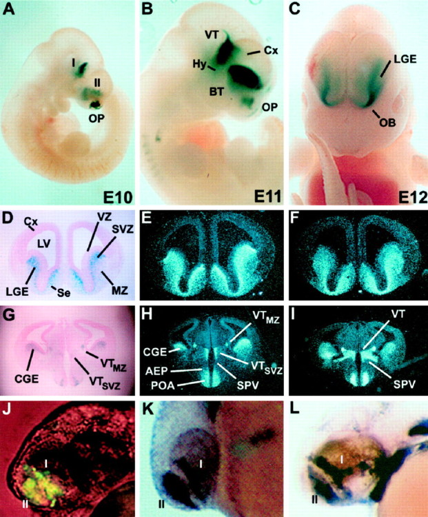
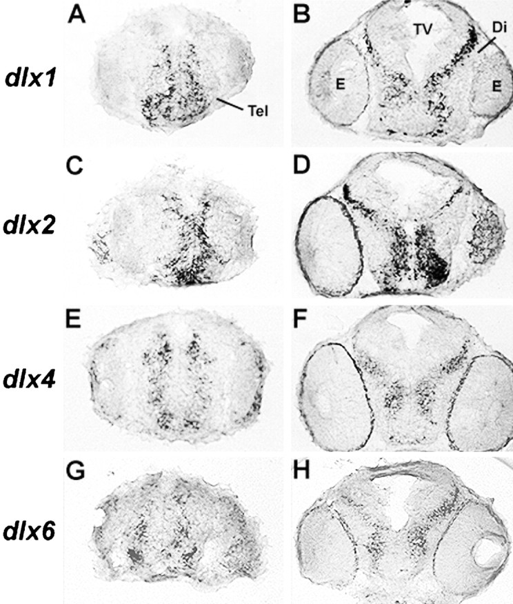
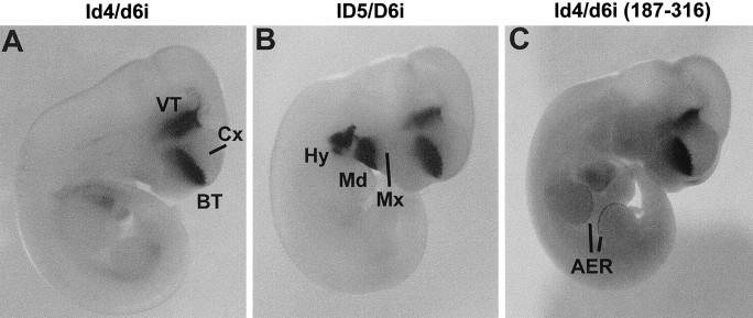
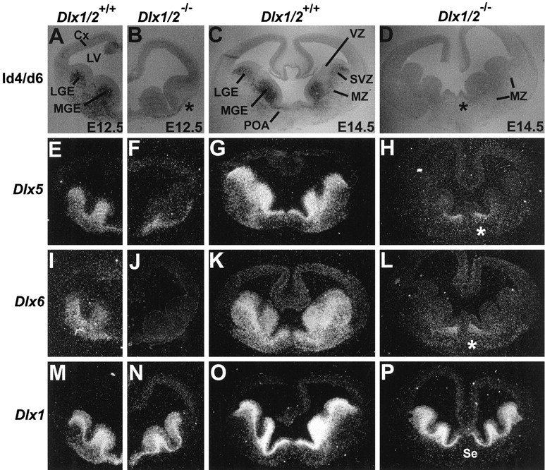
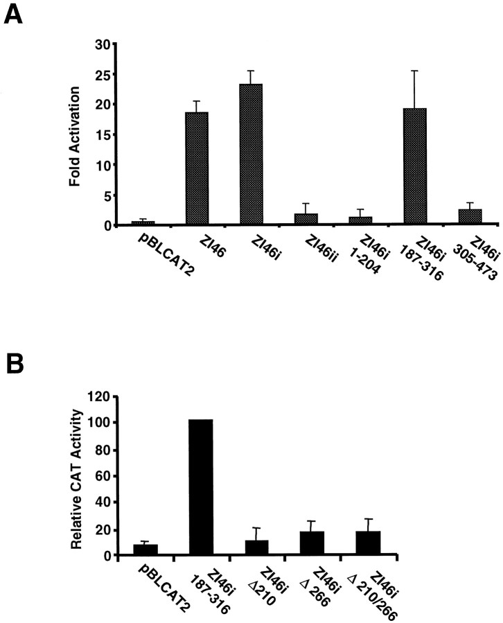
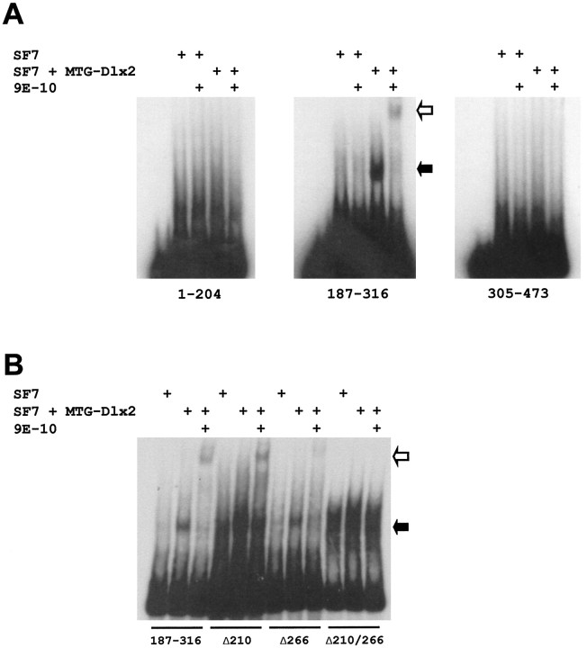
Four Dlx homeobox genes, Dlx1, Dlx2, Dlx5, and Dlx6 are expressed in the same primordia of the mouse forebrain with temporally overlapping patterns. The four genes are organized as two tail-to-tail pairs, Dlx1/Dlx2 and Dlx5/Dlx6, a genomic arrangement conserved in distantly related vertebrates like zebrafish. The Dlx5/Dlx6 intergenic region contains two sequences of a few hundred base pairs, remarkably well conserved between mouse and zebrafish. Reporter transgenes containing these two sequences are expressed in the forebrain of transgenic mice and zebrafish with patterns highly similar to endogenous Dlx5 and Dlx6 expression. The activity of the transgene is drastically reduced in mouse mutants lacking both Dlx1 and Dlx2, consistent with the decrease in endogenous Dlx5 and Dlx6 expression. These results suggest that cross-regulation by Dlx proteins, mediated by the intergenic sequences, is essential for Dlx5 and Dlx6 expression in the forebrain. This hypothesis is supported by cotransfection and DNA-protein binding experiments. We propose that the Dlx genes are part of a highly conserved developmental pathway that regulates forebrain development.


Figures

Fig. 1.
Fig. 1.
Genomic organization of the zebrafishdlx4 and dlx6 genes (top) and of the orthologous murine Dlx5 andDlx6 (bottom), indicating the location of conserved sequences with putative regulatory function. The third exons of zebrafish dlx4 and dlx6 and of mouseDlx5 and Dlx6 are represented byboxes. Direction of transcription is indicated byarrows. B, BamHI;E, EcoRI; X,XhoI; S, SacI;Sa, SalI. The constructs for the production of transgenic animals and for transfection experiments are schematized. The position and orientation of the intergenic fragments relative to the reporter genes (lacZ,CAT, or GFP) is shown.β, Minimal β-globin promoter;tk, thymidine kinase promoter. Numbers of primary transgenic embryos or embryos from transgenic lines that showlacZ expression in various sites of Dlxexpression are indicated to the right of each construct.
Fig. 2.
Fig. 2.
Conserved sequences in the intergenic region that separates a pair of vertebrate Dlx genes.A, Alignment of I56i sequences from human (h) and mouse (m) and zI46i from zebrafish (zf). B, Alignment of I56ii sequences from human and mouse and zI46ii from zebrafish. The human sequences were retrieved from GenBank, accession number AC004774. Complete sequence identity across the three species is indicated by anasterisk. The nucleotide positions of the intergenic sequences corresponding to putative homeodomain protein recognition sites as characterized by a TAAT/ATTA core sequence areshaded. The two putative Dlx binding sites that were mutagenized are shown in bold. Numbering is relative to the XhoI site (assigned the numerical position of 1) flanking a 1.4 kb element nearest to dlx6 in zebrafish and that contains both I4/6 elements (Fig. 1).
Fig. 3.
Fig. 3.
A DNA fragment encompassing the zebrafishdlx4/dlx6 intergenic region directs expression oflacZ in transgenic mouse embryos with patterns that closely recapitulate endogenous Dlx5 andDlx6 expression in the forebrain. A–C,lacZ expression in the ventral thalamus (VT), basal telencephalon (BT), and olfactory placodes (OP) in E10 (A), E11 (B), and E12 (C) whole-mount mouse embryos.D, Coronal section of an E14.5 stage mouse embryo withlacZ expression in the lateral ganglionic eminence (LGE). Higher β-galactosidase activity is seen in the subventricular zone (SVZ) compared to the mantle (MZ). E, F,In situhybridizations with Dlx5 (E) andDlx6 (F) probes on coronal sections adjacent to that seen in D. Note that the relative patterns of Dlx5 expression in the SVZ and MZ more closely resemble that seen in transgenic animals (D) than do the relative patterns ofDlx6 expression. G–I, More caudal sections of the same embryonic brain. G, Expression of β-galactosidase in the caudal ganglionic eminence (CGE), preoptic area (POA), and ventral diencephalon. H, I,In situhybridizations with Dlx5 and Dlx6 probes, respectively, on sections adjacent to those seen in G. Embryos in A and B are from line 7679, and those in C–I are from line 1469.J–L, A 1.4 kb XhoI/EcoRI fragment from the zebrafish dlx4/dlx6 intergenic region directs expression of a transgene that recapitulates endogenousdlx expression in the forebrain of zebrafish embryos.J, Expression of GFP directed by the 1.4 kb I4/6 zebrafish fragment in a 36 hr embryo. The patterns are similar to the expression of the endogenous dlx4(K) and dlx6(L) genes. The domains I and II ofdlx expression correspond, by analogy, to the diencephalic (I) and telencephalic (II) domains of Dlx expression in the mouse.I, Domain I; II, domain II;AEP, anterior entopeduncular area; BT, basal telencephalon; Cx, cortex; Hy, hypothalamus; LV, lateral ventricle; OB, prospective olfactory bulb; Se, septum;SPV, supraoptic paraventricular area; VZ, ventricular zone.
Fig. 4.
Fig. 4.
The dlx1 and dlx2genes are expressed in more immature cells of the zebrafish forebrain than their dlx4 and dlx6 paralogs. Transverse sections of 48-hr-old zebrafish embryos at the level of the telencephalon (A, C, E, G) and of the diencephalon (B, D, F, H) are shown with dorsal at thetop. Cells that express dlx1 anddlx2 are closer to the ventricle compared to those expressing dlx4 or dlx6. The expression of dlx2 closer to the ventricle compared todlx4 confirms our previous observation (Akimenko et al., 1994).
Fig. 5.
Fig. 5.
Specific intergenic sequences from either mouse or zebrafish target gene expression to the forebrain of E11 mouse embryos with highly similar patterns. A, Zebrafish zI46i.B, Mouse mI56i. In addition to the forebrain, β-galactosidase activity was observed in the first two branchial arches in two of three lines and two of four primary transgenic embryos. C, The 187–316 fragment of zebrafish zI46i. In addition to the forebrain, β-galactosidase was also expressed in the apical ectodermal ridge (AER) of the limb buds in one of five primary transgenic embryos carrying this construct. Note that the forebrain expression patterns in A–C are highly similar to those of Figure 3B (full zebrafish intergenic fragment). Mx, Maxillary component of the first branchial arch; Md, mandibular component of the first branchial arch; Hy, hyoid arch. Other abbreviations as in Figure 3.
Fig. 6.
Fig. 6.
Forebrain expression of a reporter gene driven by I4/6 is drastically reduced in mice with a targeted null mutation of the Dlx1 and Dlx2 genes. A, B, Coronal sections through the telencephalon of wild-type (A) and Dlx1/Dlx2 mutant (B) E12.5 embryos that both contain thezfdlx4/6lacZ transgene. C, D, Coronal sections through the telencephalon of wild-type (C) and mutant (D) E14.5 embryos. LacZ expression is virtually absent from the lateral (LGE) and medial (MGE) ganglionic eminences of the mutants, but is preserved in the rostral mantle (B, asterisk). E–P,In situ hybridization on sections adjacent to the ones shown inA–D with probes for Dlx5(E–H), Dlx6(I–L), and Dlx1(M–P). The Dlx1 probe recognizes a sequence in the 5′ end of the Dlx1 mRNA that is retained in the mutant (see Results). Theasterisk in D, H, andL denotes an area in the septal/preoptic region ofDlx1/Dlx2 mutants where Dlx5 andDlx6 expression at late embryonic stages appears not to be matched by lacZ expression. Other abbreviations as in Figure 3.
Fig. 7.
Fig. 7.
A, The zebrafish Dlx2 protein can activate transcription through intergenic regulatory sequences in transient transfection assays. Cotransfected Dlx2 activates transcription through the 1.4 kbXhoI/EcoRI fragment from the zebrafishdlx4/dlx6 intergenic region (I4/6), and specifically through zI46i, but not zI46ii. The 187–316 fragment from zI46i, but neither the 1–204 nor the 305–473 fragments (Fig.2A), is a target for Dlx2. All values shown represent fold activation in the presence of Dlx2 relative to the same construct in the absence of cotransfected Dlx2. B,Mutagenesis of either one of the two putative binding sites for Dlx2 in zI46i 187–316 impairs activation in transient cotransfection assays. Values shown represent the percentage of the CAT activity obtained with the wild-type zI46i 187–316 fragment. All values represent three independent experiments ± SEM.
Fig. 8.
Fig. 8.
A, The zebrafish Dlx2 protein binds the 187–316 fragment of zI46i in a gel mobility shift assay. Three fragments from zI46i: 1–204, 187–316, and 305–473 were radiolabeled and incubated with a nuclear extract from an SF7-derived cell line that expresses MTG-Dlx2 or with a control SF7 nuclear extract. A lower mobility complex is indicated by the solid arrow. In the presence of the 9E-10 antibody directed against the MTG epitope of MTG-Dlx2, this mobility complex is supershifted (open arrow). B, Mutagenesis of the two putative binding sites in the zI46i 187–316 fragment impairs binding by the Dlx2 protein. Only those lower mobility complexes obtained after incubation of MTG-Dlx2 with the wild-type 187–316 or with fragments containing one mutagenized site (Δ210 or Δ266) can be supershifted by the 9E-10 antibody. A smear around the same mobility as this retarded complex can be obtained with the fragment containing the two mutations (Δ210/266), but is also seen with the control SF7 extract and is not supershifted by the 9E-10 antibody.

Similar articles

Cited by

References

    1. Acampora D, Merlo GR, Paleari L, Zeraga B, Postiglione MP, Mantero S, Bober E, Barbieri O, Simeone A, Levi G. Craniofacial, vestibular and bone defects in mice lacking the Distal-less-related gene Dlx5. Development. 1999;126:3795–3809. - PubMed
    1. Akimenko M-A, Ekker M, Wegner J, Lin W, Westerfield M. Combinatorial expression of three zebrafish genes related to distal-less: part of a homeobox gene code for the head. J Neurosci. 1994;14:3475–3486. - PMC - PubMed
    1. Anderson SA, Eisenstat DD, Shi L, Rubenstein JLR. Interneuron migration from basal forebrain to neocortex: dependence on Dlx genes. Science. 1997a;278:474–476. - PubMed
    1. Anderson SA, Qiu M, Bulfone A, Eisenstat DD, Meneses J, Pedersen R, Rubenstein JLR. Mutations of the homeobox genes Dlx-1 and Dlx-2 disrupt the striatal subventricular zone and differentiation of late-born striatal neurons. Neuron. 1997b;19:27–37. - PubMed
    1. Andrews NC, Faller DV. A rapid micropreparation technique for extraction of DNA-binding proteins from limiting numbers of mammalian cells. Nucleic Acids Res. 1991;19:2499. - PMC - PubMed

Publication types