. 2000 Jan 15;20(2):806-12.
doi: 10.1523/JNEUROSCI.20-02-00806.2000.

GABA(C) receptors control adaptive changes in a glycinergic inhibitory pathway in salamander retina

Affiliations

GABA(C) receptors control adaptive changes in a glycinergic inhibitory pathway in salamander retina

P B Cook et al. J Neurosci. .

Abstract

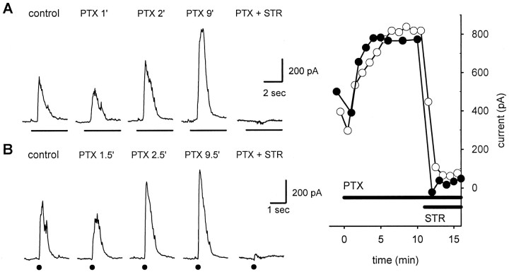
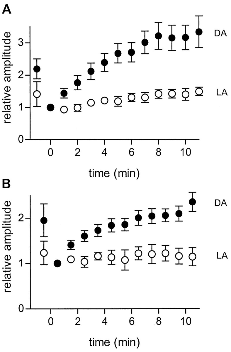
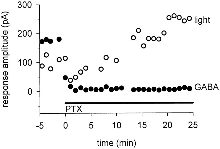
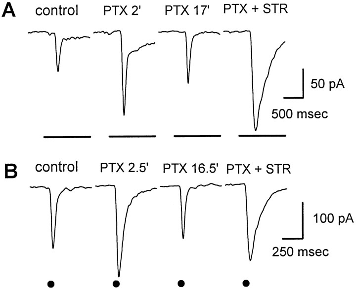
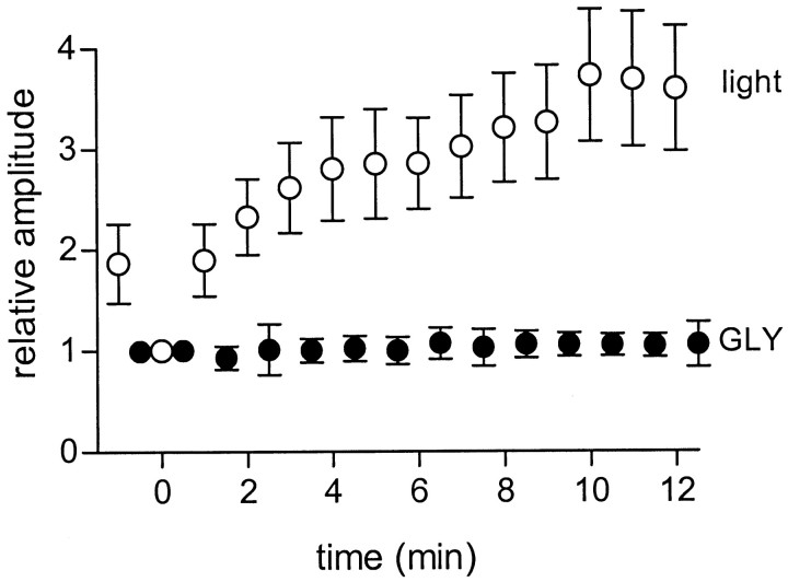
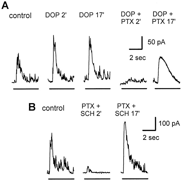
We studied the role of GABA in adaptive changes in a lateral inhibitory system in the tiger salamander retina. In dark-adapted retinal slice preparations picrotoxin caused a slow enhancement of glycine-mediated IPSCs in ganglion cells. The enhancement of glycinergic IPSCs developed slowly over the course of 5-20 min, even though picrotoxin blocked both GABA(A) and GABA(C) receptors within a few seconds. The slow enhancement of glycinergic IPSCs by picrotoxin was much weaker in light-adapted preparations. The slow enhancement of glycinergic inhibitory inputs was not produced by bicuculline, indicating that it involved GABA(C) receptors. The responses of ganglion cells to direct application of glycine were not enhanced by picrotoxin, indicating that the enhancement was not caused by an action on glycine receptors. In dark-adapted eyecup preparations picrotoxin caused a slow enhancement of glycinergic IPSPs and transient lateral inhibition produced by a rotating windmill pattern, similar to the effect of light adaptation. The results suggest that the glycinergic inhibitory inputs are modulated by an unknown substance whose synthesis and/or release is inhibited in dark-adapted retinas by GABA acting at GABA(C) receptors.


Figures

Fig. 1.
Fig. 1.
Effects of picrotoxin on inhibitory currents in ganglion cells. Responses are from an on–off ganglion cell in a tiger salamander slice preparation. The cell was voltage clamped at 0 mV to eliminate glutamate-mediated excitatory currents.A, IPSCs elicited by 4 sec full-field light stimuli (indicated by the horizontalbarbeloweachtrace).Traces show IPSCs recorded before application of picrotoxin (control) and 1, 2, and 9 min after onset of superfusion with picrotoxin (PTX 1′, 2′, and9′). Subsequent addition of 2 μmstrychnine in the continued presence of picrotoxin (PTX + STR) completely blocked the enhanced IPSCs, indicating that they were mediated by glycine. The holding current was +42 pA. The light flashes also elicited IPSCs at light off (data not shown); the effects on the off responses were similar to those of the on responses.B, Same as A, except that the IPSCs were evoked by zaps (+0.5 μA; 1 msec) in the outer plexiform layer directly above the recorded ganglion cell. The time of the zap stimulus is indicated by dotbelow each response.Inset,Right, Peak light-evoked (filledcircles) and zap-evoked (opencircles) IPSC amplitudes at additional times.
Fig. 2.
Fig. 2.
Enhancement of glycinergic IPSCs by picrotoxin is greater in dark- than light-adapted retinas. A, IPSCs elicited by full-field light flashes at different times after onset of continuous superfusion with picrotoxin. Data are averaged from 16 dark-adapted retinas (filledcircles) and 7 light-adapted retinas (opencircles). For each cell, peak IPSC amplitudes were normalized to that of the first response recorded in the presence of picrotoxin, which was recorded within 1 min after switching the superfusate to picrotoxin. Superfusion with picrotoxin begins at t = 0. In 11 of the cells tested (8 dark-adapted, 3 light-adapted) 2 μm strychnine was added after the enhancement had reached a maximum; in all cases strychnine completely blocked the IPSCs, indicating that they were glycinergic. Error bars indicate 1 SEM. The maximum enhancement was 3.57 (± 0.33)-fold in dark-adapted preparations (n = 16) and 1.49 (± 0.15)-fold in light-adapted preparations (n = 7). The probability that this difference was caused by chance was < 0.003 (Student's unpairedt test). B, Same as A, except that the IPSCs were elicited by focal electrical stimuli (+0.5 μA; 1 msec) as described in Figure 1. The maximum enhancement was 2.38 (± 0.20)-fold for dark-adapted preparations (n = 4) and 1.12 (± 0.28)-fold for light-adapted preparations (n = 4). The probability that this difference was caused by chance was < 0.001 (Student's unpaired t test). DA, Dark-adapted; LA, light-adapted.
Fig. 3.
Fig. 3.
Effect of picrotoxin on ganglion cell IPSCs in a light-adapted retinal slice. Left, Details in Figure 1, except that this retina had been light adapted by exposure to background illumination for several minutes. Inset,Right, Superimposed traces of the first two responses (control and PTX 1′) scaled to the same amplitude to better illustrate the difference in time courses.
Fig. 4.
Fig. 4.
Picrotoxin slowly enhances light-evoked glycinergic responses but rapidly blocks responses to directly applied GABA. Responses are IPSCs evoked by alternating full-field light flashes (opencircles) and puffs of GABA (filledcircles). Picrotoxin (PTX) rapidly blocked the responses to GABA puffs, but the light-evoked IPSCs were initially reduced and then slowly enhanced over the next 20 min. Similar results were seen in all of the four cells tested.
Fig. 5.
Fig. 5.
Bicuculline does not enhance glycinergic IPSCs in ganglion cells. A, Light-evoked IPSCs.Opencircles show the amplitude of IPSCs at various times after onset of continuous superfusion with 200 μm bicuculline (BIC). For comparison,filledcircles show the results (from Fig. 2A) obtained during superfusion with 150 μm picrotoxin (PTX). Data were averaged from 16 cells for PTX and 5 cells forBIC. For each cell, peak IPSC amplitudes were normalized to the first response recorded in the presence of picrotoxin.B, Same as A, except that the IPSCs were elicited by zaps (+0.5 μA; 1 msec) as described in Figure 1.
Fig. 6.
Fig. 6.
Effect of picrotoxin on excitatory currents in an on–off ganglion cell. The cell was voltage clamped at the chloride reversal potential (−65 mV) to eliminate GABA- and glycine-mediated inhibitory currents. Other details are as described in Figure 1.A, EPSCs that were elicited by 4 sec full-field light stimuli. After the onset of picrotoxin the EPSCs were initially enhanced (PTX 2′) but then gradually declined (PTX 17′). Subsequent addition of 2 μmstrychnine in the continued presence of picrotoxin (PTX + STR) partially reversed the slow decline in EPSC amplitude caused by picrotoxin, indicating that the slowly developing suppression of the EPSC was mediated by glycine. The holding current was −52 pA. The effects on the off responses (data not shown) were similar.B, Same as A, except that the EPSCs were elicited by zaps (+0.5 μA; 1 msec) as described in Figure 1.
Fig. 7.
Fig. 7.
Picrotoxin does not affect the ganglion cell response to direct application of glycine. Datapoints indicate the average peak amplitude of IPSCs elicited by full-field illumination (opencircles) and puffs of glycine (filledcircles) in four cells at various times after onset of continuous superfusion with picrotoxin att = 0. For each cell, responses were normalized to the first response after the application of picrotoxin in each cell.GLY, Glycine.
Fig. 8.
Fig. 8.
Effect of dopamine and dopamine antagonists on the ability of picrotoxin to cause slow enhancement of glycinergic IPSCs. Light stimuli and recording conditions are as described in Figure 1.A, Addition of 20 μm dopamine caused a rapid increase in IPSC amplitude within 2 min (DOP 2′), but there was no further increase after 15 additional minutes in dopamine (DOP 17′). The initial enhancement of the IPSC by dopamine was seen in only two of the four cells tested; in all four cells the mean enhancement was 1.13 (± 0.32)-fold (p = 0.65). After 18 min in dopamine, addition of 150 μm picrotoxin caused an immediate strong reduction in the IPSC (DOP + PTX 2′). In the continued presence of PTX the IPSC was slowly enhanced (DOP + PTX 17′). The mean enhancement by PTX was 4.02 (± 1.48)-fold (p = 0.02;n = 4). B, Addition of 15 μm SCH23390 and 150 μmPTXcaused an immediate reduction of the IPSC (SCH + PTX 2′), but in the continued presence of PTX andSCH the response slowly became enhanced (SCH + PTX 17′). In the five cells tested, the maximum enhancement byPTX in the presence of SCH was 4.42 (± 1.24)-fold (p = 0.02). In bothA and B the enhanced IPSC inPTX was completely blocked by 2 μmstrychnine in all cells (data not shown).
Fig. 9.
Fig. 9.
Effect of picrotoxin on transient lateral inhibition and glycinergic IPSPs in on–off ganglion cells in eyecup preparations. A, Effect of 150 μmpicrotoxin (PTX) on TLI in a dark-adapted eyecup. TLI was measured as the suppression of the response to a small test spot in the receptive field center by rotation of a broken annulus (windmill) pattern. B, Effect of picrotoxin on the transient IPSP elicited at the onset of an annulus (i.d., 500 μm; o.d., 2600 μm) in a light-adapted eyecup. Traces show the IPSP response in control Ringer's solution and at 2 and 10 min after onset of continuous superfusion with 150 μmpicrotoxin. The horizontallinebelow the responsetracesindicates the initial portion of the 4 sec light stimulus.

Similar articles

Cited by

References

    1. Belgum JH, Dvorak DR, McReynolds JS. Strychnine blocks transient but not sustained inhibition in mudpuppy retinal ganglion cells. J Physiol (Lond) 1984;354:273–286. - PMC - PubMed
    1. Cook PB, McReynolds JS. Modulation of sustained and transient lateral inhibitory mechanisms in the mudpuppy retina during light adaptation. J Neurophysiol. 1998;79:197–204. - PubMed
    1. Cook PB, Lukasiewicz PD, McReynolds JS. Action potentials are required for the lateral transmission of glycinergic transient inhibition in the amphibian retina. J Neurosci. 1998;18:2301–2308. - PMC - PubMed
    1. Dong CJ, Werblin FS. Temporal contrast enhancement via GABAC feedback at bipolar terminals in the tiger salamander retina. J Neurophysiol. 1998;79:2171–2180. - PubMed
    1. Dowling JE. Retinal neuromodulation: the role of dopamine. Vis Neurosci. 1991;1:87–97. - PubMed

Publication types

LinkOut - more resources