. 2000 Jan 15;20(2):820-33.
doi: 10.1523/JNEUROSCI.20-02-00820.2000.

Relationship of activity in the subthalamic nucleus-globus pallidus network to cortical electroencephalogram

Affiliations

Relationship of activity in the subthalamic nucleus-globus pallidus network to cortical electroencephalogram

P J Magill et al. J Neurosci. .

Abstract

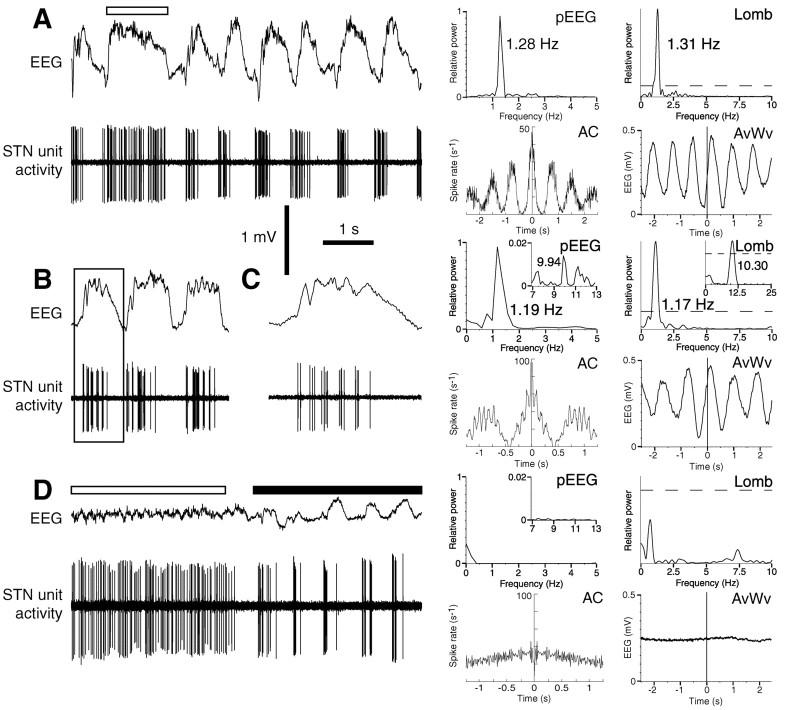
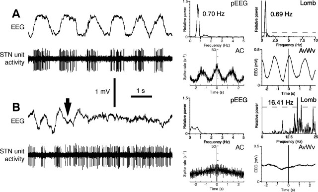
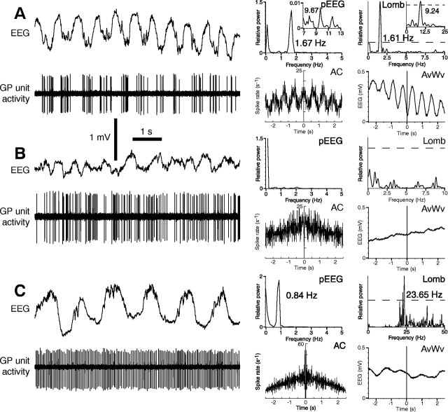
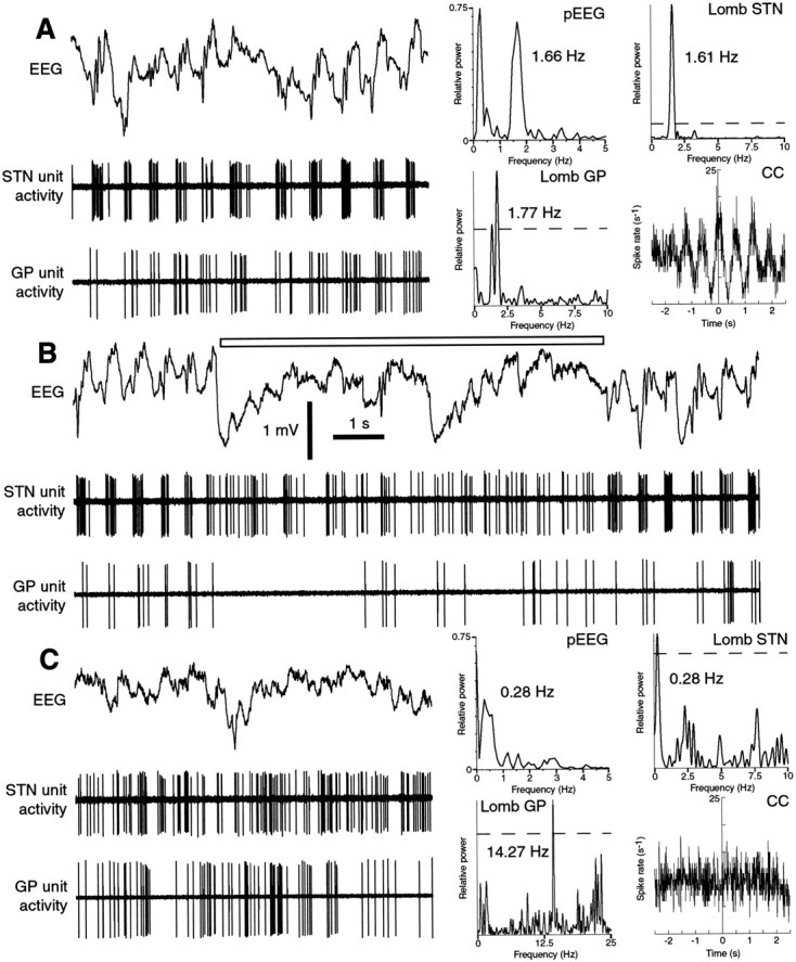
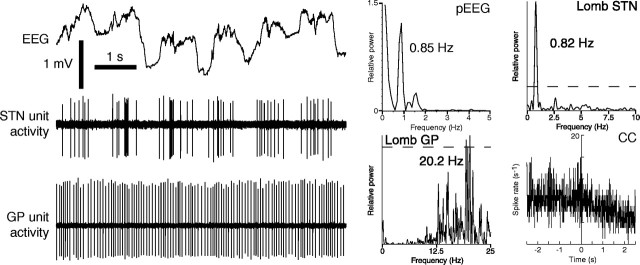
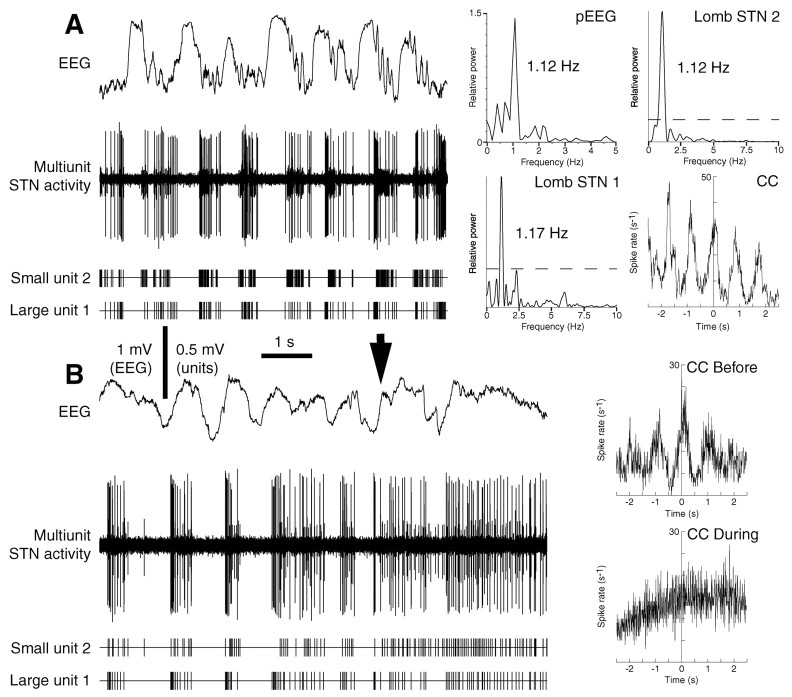
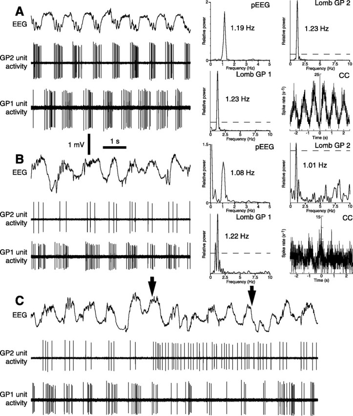
One of the functions of the excitatory subthalamic nucleus (STN) is to relay cortical activity to other basal ganglia structures. The response of the STN to cortical input is shaped by inhibition from the reciprocally connected globus pallidus (GP). To examine the activity in the STN-GP network in relation to cortical activity, we recorded single and multiple unit activity in STN and/or GP together with cortical electroencephalogram in anesthetized rats during various states of cortical activation. During cortical slow-wave activity (SWA), STN and GP neurons fired bursts of action potentials at frequencies that were similar to those of coincident slow ( approximately 1 Hz) and spindle (7-14 Hz) cortical oscillations. Spontaneous or sensory-driven global activation was associated with a reduction of SWA and a shift in STN-GP activity from burst- to tonic- or irregular-firing. Rhythmic activity in STN and GP neurons was lost when the cortex was inactivated by spreading depression and did not resume until SWA had recovered. Although rhythmic STN-GP activity was correlated with SWA, the phase relationships of activities of neurons within the STN and GP and between the nuclei were variable. Even when neurons displayed synchronous bursting activity, correlations on the millisecond time scale, which might indicate shared synaptic input, were not observed. These data indicate that (1) STN and GP activity is intimately related to cortical activity and hence the sleep-wake cycle; (2) rhythmic oscillatory activity in the STN-GP network in disease states may be driven by the cortex; and (3) activity of the STN-GP network is regulated in space in a complex manner.


Figures

Fig. 1.
Fig. 1.
Light micrographs of subthalamic nucleus and globus pallidus neurons that were juxtacellularly labeled with neurobiotin. A, This STN neuron was located in the caudal portion of the more darkly stained STN. ZIV, Ventral division of the zona incerta; CP, cerebral peduncle. B, This GP neuron was situated in the rostral aspect of the GP. A blood vessel (∗) lies on the border between the GP and the more darkly stained neostriatum (NS). Rostral is to the left, and dorsal is to the topof each figure. Scale bars, 200 μm.
Fig. 2.
Fig. 2.
Spike-firing patterns of subthalamic nucleus neurons are related to coincident cortical activity during ketamine anesthesia. A, This STN neuron had a BI of 1.43 and fired robust bursts of spikes on the rising phase of the slow cortical oscillation. Note that during periods of prolonged cortical activation (under white bar), the burst duration was increased. Rhythmic spike-firing was manifest as peaks in the autocorrelogram (AC). Comparison of the Lomb periodogram (Lomb) with the power spectrum of the EEG (pEEG) shows a similar frequency of rhythmic activity in the STN spike train and cortex. Dashed linein this and subsequent Lomb periodograms denotes the significance level of p = 0.05. The phase relationship between spiking and the EEG waves is shown on the spike-triggered waveform average (AvWv). B, The firing of another bursty neuron (BI = 1.0) was phase-locked to the crest of the cortical slow-wave. Note the smaller amplitude, spindle-like events (frequency ∼10 Hz) superimposed on the peaks of the slow-wave in the EEG trace. The large bursts occurred at a frequency of ∼1 Hz and were composed of a number of “miniature bursts,” which are shown as small peaks riding on the top of the three larger peaks in the autocorrelogram. The main Lomb periodogram shows significant bursting at a frequency very similar to that of the large slow-wave. The inset Lomb is filtered between 4 and 25 Hz and shows a significant oscillation in the spike train at ∼10 Hz frequency. C, Theboxed area in B (1 sec of data) on an expanded time scale. The firing of miniature bursts was phase-locked with the generation of the spindle-like wavelets. D, Burst activity in the same neuron was replaced with irregular, single-spike activity during episodes of cortical spreading depression (white bar). The four graphs to the rightof the trace in D were constructed from the period under the white bar; note that a significant oscillation in the Lomb was no longer present and that the autocorrelogram did not have peaks when power in the EEG was severely attenuated. Partial recovery of cortical SWA was accompanied by a partial restoration of burst activity (black bar). Calibration bars apply to all panels except C. In this and the following figures, AC designates autocorrelograms of spiking activity (bin size 10 msec), Lomb designates Lomb periodograms of spiking activity, pEEG designates power spectra of the coincident EEG, and AvWv designates spike-triggered averages of EEG.
Fig. 3.
Fig. 3.
Spike-firing patterns of subthalamic nucleus neurons are related to coincident cortical activity in urethane anesthesia. A, A bursty STN neuron (BI = 0.67), the firing of which was phase-locked to SWA. Note the significantly lower frequency of SWA and periodic bursting as compared with activity during ketamine anesthesia. B, Disruption of SWA by sensory stimulation (hindpaw pinch of 10 sec duration; starts at black arrow) was concomitant with a loss of bursty activity in the same neuron. AC, Lomb, pEEG, and AvWv in B were determined during the pinch. Calibration bars apply to both panels.
Fig. 4.
Fig. 4.
Spike-firing patterns of globus pallidus neurons are related to coincident cortical activity. A, A bursty, pallidostriatal GP neuron (BI = 0.67) firing well defined bursts of spikes during the troughs of the slow-wave during ketamine anesthesia. Comparison of the Lomb with the power spectrum shows very similar frequencies of rhythmic activity in the spike train and EEG. The main Lomb periodogram shows significant bursting at a frequency very similar to that of the large slow-wave; the insetLomb is filtered between 4 and 25 Hz and shows a significant oscillation in the spike train at ∼10 Hz frequency, which is similar to the spindle frequency. B, During cortical spreading depression, the amplitude of the SWA was attenuated, and burst activity in the same neuron was replaced with irregular, single-spike activity. This neuron eventually became quiescent after prolonged depression of cortical activity. The neuron did not burst again until cortical SWA had recovered. C, During urethane anesthesia, GP neurons displayed a highly regular, single-spike firing pattern that was persistent during episodes of robust slow-wave activity. The GP neuron in C had a mean firing rate of 23.7 Hz, which is similar to the dominant frequency of activity in the Lomb, thus confirming the regular, tonic nature of its spiking. Calibration bars apply to all panels.
Fig. 5.
Fig. 5.
Simultaneous recordings of a subthalamic nucleus neuron and a globus pallidus neuron during ketamine anesthesia.A, This pair of bursty neurons displayed near synchronous bursting during robust SWA. Burst indices of the STN and GP neurons were 1.43 and 0.50, respectively. Burst firing of both neurons was phase-locked to the slow cortical oscillation. The cross-correlogram (CC) possessed several broad peaks: on average, the STN neuron fired ∼30 msec before the GP neuron (18° phase difference). Narrow peaks on the millisecond time scale were not observed, which implies that the pair were not monosynaptically connected. B, Spontaneous, brief periods of reduction of slow-wave amplitude and rhythmicitywere associated with a loss of correlated activity in the same neurons (under white bar). Correlated burst-firing swiftly resumed when robust SWA was restored. C, Later in the same recording session, SWA effectively collapsed for ∼40 sec, and periodicity and correlation were lost in the pair. Note that the GP neuron fired faster during the prolonged loss of SWA inC as compared with the short-lived loss of SWA inB. In this and the following figures, CCdesignates cross-correlograms of spiking activity between pairs of neurons (bin size 10 msec). Calibration bars apply to all panels.
Fig. 6.
Fig. 6.
Simultaneous recordings of a subthalamic nucleus neuron and a globus pallidus neuron during urethane anesthesia.A, Typical example of uncorrelated firing in the STN–GP network. Although STN neurons fired bursts of spikes in a discrete, phase-locked manner, all GP neurons maintained a regular firing mode under this anesthetic regimen.
Fig. 7.
Fig. 7.
Near neighbors in the subthalamic nucleus exhibit synchronous, phase-locked firing during robust SWA. A, Multiunit recording from a single electrode during ketamine anesthesia demonstrated that the firing of STN neurons in close proximity was tightly correlated. This pair showed a small phase lag of ∼30 msec (12°). The bottom two traces are spike-triggered digital-pulse trains dissected from the unit recordings and show more clearly the individual patterns of spike-firing of the two neurons. Both neurons had a BI of 1.43. B, Multiunit recordings showing near synchronous firing of neighboring STN neurons during urethane anesthesia (phase difference of ∼60 msec). Disruption of the robust SWA by pinching (black arrow) was associated with transitions to irregular single-spike firing by the two neurons and a loss of correlated activity. Calibration bars apply to both panels.
Fig. 8.
Fig. 8.
Simultaneous recordings of pairs of globus pallidus neurons during ketamine anesthesia. A, Phase-locked burst-firing in a pair of GP neurons. The neurons had an identical frequency of oscillation in their spike trains, which was similar in frequency to the cortical slow oscillation. Unit 1 (BI = 0.5) selectively fired during the inactive component of the SWA, and unit 2 (BI = 1.0) only fired during the active component. Thus, their firing was inversely correlated with a phase lag of ∼400 msec (176°). B, The burst-firing of another pair of GP neurons was much more closely synchronized with a phase difference of ∼60 msec (24°). C, Heterogeneous response of neurons in B to a 5 sec hindpaw pinch (between black arrows). Unit 2 adopted a regular-firing mode with a small increase in firing rate; in contrast, unit 1 maintained a burst-firing mode, but with a decreased firing rate. This diverse response resulted in uncorrelated firing between the two neurons. Calibration bars apply to all panels.

Similar articles

Cited by

References

    1. Abeles M. Quantification, smoothing, and confidence limits for single-units' histograms. J Neurosci Methods. 1982;5:317–325. - PubMed
    1. Achermann P, Borbély AA. Low-frequency (<1 Hz) oscillations in the human sleep electroencephalogram. Neuroscience. 1997;81:213–222. - PubMed
    1. Adrian ED, Matthews BHC. The interpretation of potential waves in the cortex. J Physiol (Lond) 1934;81:440–471. - PMC - PubMed
    1. Albe-Fessard D, Condes-Lara M, Kesar S, Sanderson P. Tonic cortical controls acting on spontaneous and evoked thalamic activity. In: Macchi G, Rustioni A, Spreafico R, editors. Somatosensory integration in the thalamus. Elsevier; Amsterdam: 1983. pp. 273–285.
    1. Aldridge JW, Gilman S. The temporal structure of spike trains in the primate basal ganglia: afferent regulation of bursting demonstrated with precentral cerebral cortical ablation. Brain Res. 1991;543:123–138. - PubMed

Publication types

LinkOut - more resources