

Development of dextran sulphate sodium-induced experimental colitis is suppressed in genetically mast cell-deficient Ws/Ws rats
- PMID: 10632661
- PMCID: PMC1905515
- DOI: 10.1046/j.1365-2249.2000.01094.x
Development of dextran sulphate sodium-induced experimental colitis is suppressed in genetically mast cell-deficient Ws/Ws rats
Abstract
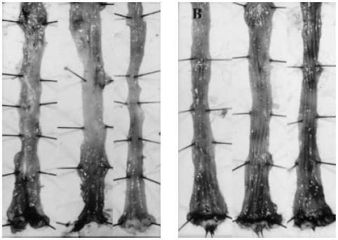
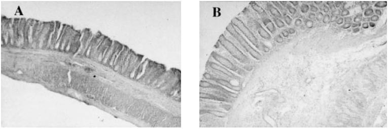
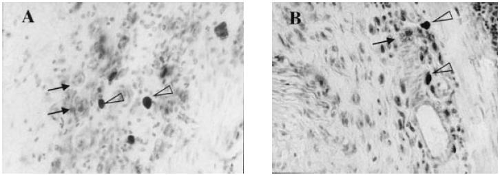
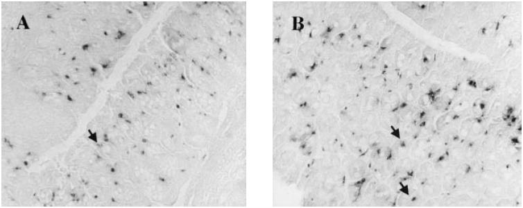
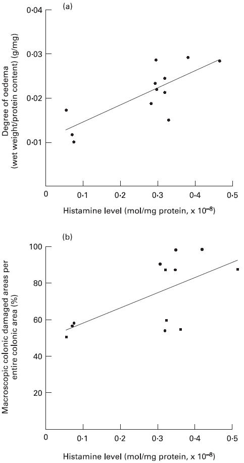
Ws/Ws rats have a small deletion of the c-kit gene, and are deficient in both mucosal-type mast cells (MMC) and connective tissue-type mast cells (CTMC). In the present study we investigated the role of intestinal MMC in the development of dextran sulphate sodium (DSS)-induced experimental colitis using Ws/Ws rats. Ws/Ws and control (+/+) rats were given a 3% DSS aqueous solution orally for 10 days, and the subsequent mucosal damage was evaluated macroscopically and histologically. The mucosal myeloperoxidase (MPO) activities and histamine levels were also measured. (i) DSS induced severe oedema and hyperaemia with sporadic erosions in the control (+/+) rats, but these changes were significantly attenuated in the Ws/Ws rats (P < 0.01). (ii) The microscopic mucosal damage score was lower in the Ws/Ws rats than in the control (+/+) rats (P = 0.06). (iii) There were no significant differences in mucosal MPO activity between the Ws/Ws and control (+/+) rats (P = 0.46). (iv) The mucosal histamine levels in the colon were significantly reduced in the Ws/Ws rats compared with the control (+/+) rats (P < 0.05). (v) Significant positive correlations were observed between mucosal histamine levels and the degree of mucosal oedema (calculated as colonic wet weight/protein content) (r = 0.778, P < 0.01), and between histamine levels and the macroscopic damage (r = 0.623, P < 0.05), respectively. (vi) DSS induced a local recruitment of MMC in the colonic mucosa of Ws/Ws rats, and mucosal damage gradually increased in accordance with this MMC recruitment. These results indicate that MMC play an important role in the development of DSS colitis.
Figures
References
-
- Boros M, Kaszaki J, Nagy S. Histamine release during intestinal ischemia–reperfusion: role of irons and hydrogen peroxide. Circ Shock. 1991;35:174–80. - PubMed
-
- Boros M, Kaszaki J, Nagy S. Oxygen free radical induced histamine release during intestinal ischemia and reperfusion. Eur Surg Res. 1989;21:297–304. - PubMed
-
- Kubes P, Ibbotson G, Russell JM, et al. Role of platelet-activating factor in ischemia/reperfusion induced leukocyte adherence. Am J Physiol. 1990;259:G300–5. - PubMed
-
- McAuley RL, Somers SC. Mast cells in nonspecific ulcerative colitis. Am J Dig Dis. 1961;6:233–6.
MeSH terms
Substances
LinkOut - more resources
Full Text Sources
Other Literature Sources
Medical
Research Materials
Miscellaneous
