

Changes in the cytokine profile of lupus-prone mice (NZB/NZW)F1 induced by Plasmodium chabaudi and their implications in the reversal of clinical symptoms
- PMID: 10632672
- PMCID: PMC1905516
- DOI: 10.1046/j.1365-2249.2000.01124.x
Changes in the cytokine profile of lupus-prone mice (NZB/NZW)F1 induced by Plasmodium chabaudi and their implications in the reversal of clinical symptoms
Abstract
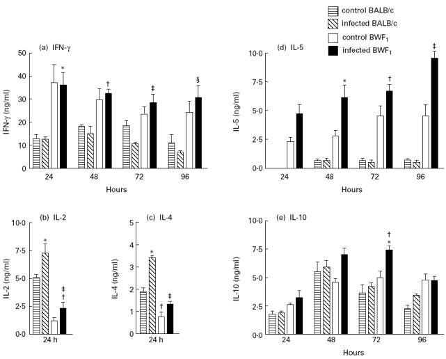
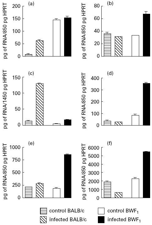
We have previously observed that aged lupus-prone (NZB/NZW)Fl (BWF1) mice when infected with Plasmodium chabaudi show an improvement in their clinical lupus-like symptoms. In order to study the mechanisms involved in the long-lasting protective effect of the P. chabaudi infection in lupus-prone mice we analysed specific aspects of the cellular response, namely the profiles of cytokine mRNA expression and cytokine secretion levels in old BWF1 mice, in comparison with uninfected age-matched BWF1 mice and infected or uninfected BALB/c mice. Two months after infection, cells from BWF1 mice were stimulated with concanavalin A (Con A) and demonstrated a recovery of T cell responsiveness that reached the levels obtained with BALB/c cells. Old BWF1 mice showed high levels of interferon-gamma (IFN-gamma) and IL-5 production and correspondingly low levels of IL-2 and IL-4 secretion before infection with P. chabaudi. Infection did not modify the IFN-gamma levels of BWF1 T cells, whereas it considerably increased the secretion of the Th2-related cytokines IL-4, IL-5 and IL-10. In addition, only BWF1 T cells showed increased mRNA expression of tumour necrosis factor-alpha (TNF-alpha) and transforming growth factor-beta (TGF-beta). This counter-regulatory cytokine network of infected BWF1 mice may be involved in the improvement of their lupus symptoms. The results of our investigations using the complex model of P. chabaudi infection can be extended and, by using more restricted approaches, it may be possible to explain the multiple regulatory defects of lupus-prone mice.
Figures
Similar articles
-
Differences between CD8+ T cells in lupus-prone (NZB x NZW) F1 mice and healthy (BALB/c x NZW) F1 mice may influence autoimmunity in the lupus model.Eur J Immunol. 2004 Sep;34(9):2489-99. doi: 10.1002/eji.200424978. Eur J Immunol. 2004. PMID: 15307181 Free PMC article.
-
Defects in the regulation of anti-DNA antibody production in aged lupus-prone (NZB x NZW)F1 mice: analysis of T-cell lymphokine synthesis.Immunology. 1995 May;85(1):26-32. Immunology. 1995. PMID: 7635519 Free PMC article.
-
Ifi202, an IFN-inducible candidate gene for lupus susceptibility in NZB/W F1 mice, is a positive regulator for NF-kappaB activation in dendritic cells.Int Immunol. 2007 Aug;19(8):935-42. doi: 10.1093/intimm/dxm054. Epub 2007 Aug 16. Int Immunol. 2007. PMID: 17702989
-
Dendritic cells, pro-inflammatory responses, and antigen presentation in a rodent malaria infection.Immunol Rev. 2004 Oct;201:35-47. doi: 10.1111/j.0105-2896.2004.00182.x. Immunol Rev. 2004. PMID: 15361231 Review.
-
Aberrant B1 cell trafficking in a murine model for lupus.Front Biosci. 2007 Jan 1;12:1790-803. doi: 10.2741/2188. Front Biosci. 2007. PMID: 17127421 Review.
Cited by
-
SLE: Another Autoimmune Disorder Influenced by Microbes and Diet?Front Immunol. 2015 Nov 30;6:608. doi: 10.3389/fimmu.2015.00608. eCollection 2015. Front Immunol. 2015. PMID: 26648937 Free PMC article. Review.
-
Glomerulonephritis, Th1 and Th2: what's new?Clin Exp Immunol. 2005 Nov;142(2):207-15. doi: 10.1111/j.1365-2249.2005.02842.x. Clin Exp Immunol. 2005. PMID: 16232206 Free PMC article. Review.
-
Down-regulation of pathogenic autoantibody response in a slowly progressive Heymann nephritis kidney disease model.Int J Exp Pathol. 2004 Dec;85(6):321-34. doi: 10.1111/j.0959-9673.2004.00388.x. Int J Exp Pathol. 2004. PMID: 15566429 Free PMC article.
-
The role of dendritic cells in the mechanism of action of a peptide that ameliorates lupus in murine models.Immunology. 2009 Sep;128(1 Suppl):e395-405. doi: 10.1111/j.1365-2567.2008.02988.x. Epub 2008 Nov 24. Immunology. 2009. PMID: 19040426 Free PMC article.
References
-
- Theofilopoulos AN, Dixon J. Murine models of systemic lupus erythematosus. Adv Immunol. 1985;37:269–390. - PubMed
-
- Merino RM, Iwamoto M, Fossati L, Izui S. Polyclonal B cell activation arises from different mechanisms in lupus-prone (NZB × NZW) F1 and MRL/MpJ-Ipr/Ipr mice. J Immunol. 1993;152:6509–16. - PubMed
-
- Hentati B, Ternynck T, Avrameas S, Ternynck T. Comparison of natural antibodies to autoantibodies arising during lupus in (NZB × NZW) F1 mice. J Autoimmun. 1991;4:341–56. - PubMed
-
- Greenwood BM, Herrick EM, Voller A. Suppression of autoimmune disease in NZB and (NZW × NZB) F1 hybrid mice by infection with malaria. Nature. 1970;226:266–7. - PubMed
Publication types
MeSH terms
Substances
LinkOut - more resources
Full Text Sources
Medical
Molecular Biology Databases
