. 2000 Feb;119(2):361-7.
doi: 10.1046/j.1365-2249.2000.01125.x.

Exacerbation of acute inflammatory arthritis by the colony-stimulating factors CSF-1 and granulocyte macrophage (GM)-CSF: evidence of macrophage infiltration and local proliferation

Affiliations

Exacerbation of acute inflammatory arthritis by the colony-stimulating factors CSF-1 and granulocyte macrophage (GM)-CSF: evidence of macrophage infiltration and local proliferation

R J Bischof et al. Clin Exp Immunol. 2000 Feb.

Abstract

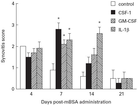
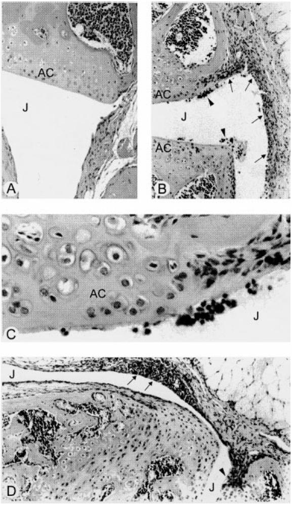
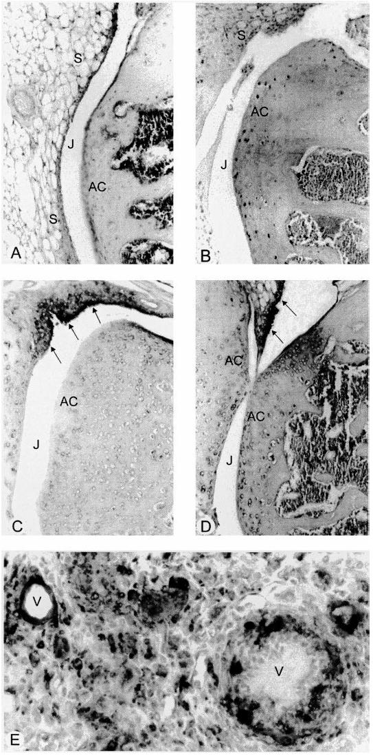
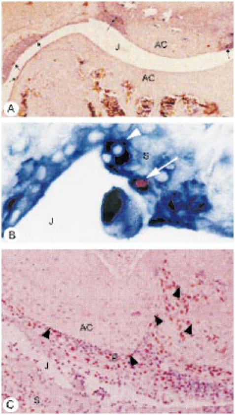
CSF-1 and GM-CSF have been implicated in the pathogenesis of rheumatoid arthritis. We report the effects of CSF-1 and GM-CSF in the development of an acute methylated bovine serum albumin (mBSA)-induced murine arthritis model. Examination of histopathological features revealed that the systemic administration of CSF-1 or GM-CSF following mBSA administration into the knee resulted in the exacerbation of arthritis. This included synovial hyperplasia and joint inflammation, most evident at 7 and 14 days post-mBSA administration, and the appearance of erosive pannus tissue. The exacerbation by CSF-1 and GM-CSF was not sustained but declined in incidence and severity by 21 days post-mBSA administration, similar to the effects of IL-1beta in this model, reported here and previously. Macrophages expressing Mac-2 and F4/80 were a prominent feature of the pathology observed, particularly the infiltration of Mac-2+ macrophages seen in all mice administered CSF-1, GM-CSF or IL-1beta. Present in inflamed knees was a locally dividing population of cells which included Mac-2+ and F4/80+ macrophages. These studies demonstrate that CSF-1 and GM-CSF can exacerbate and prolong the histopathology of acute inflammatory arthritis and lend support to monocytes/macrophages being a driving influence in the pathogenesis of inflammatory arthritis.


Figures

Fig. 1
Fig. 1
Histopathological scores for synovial hyperplasia in methylated bovine serum albumin (mBSA)-injected knees. Mice were given an i.a. injection of mBSA on day 0 of the experiment together with three daily s.c. injections (days 0–2) of either saline (control), CSF-1 (7.5 μg), granulocyte macrophage (GM)-CSF (15 μg) or IL-1β (250 ng). The severity of synovial hyperplasia was scored on a scale of 0 (normal) to 3 (severe) (see Materials and Methods) following sacrifice on day 4, 7, 14 or 21 of the experiment. Values are presented as mean scores (± s.e.m.) obtained from groups of 10 mice. *P < 0.05 compared with the mBSA/saline control group; Mann–Whitney two-sample rank test.
Fig. 2
Fig. 2
Histopathological manifestations of acute arthritis 7 days after administration of methylated bovine serum albumin (mBSA) and either CSF-1, granulocyte macrophage (GM)-CSF or IL-1β. (A) Relatively normal (non-arthritic) joint (frontal knee section) from mouse administered mBSA/saline showing normal joint architecture and no evidence of inflammation. (B) Representative frontal knee joint section from an mBSA/GM-CSF-injected mouse showing a thickened, inflamed synovium (arrows) and inflammatory cells (arrowheads) in the joint space. (C) Higher magnification of area in (B) showing detail of the inflammatory exudate, typically comprising monocytes/macrophages and neutrophils, some seen in contact with the articular cartilage and synovium. (D) Inflammatory cells localized around some of the larger blood vessels (arrows) of the inflamed synovium (in this instance taken from a mouse injected with mBSA/CSF-1). Note also invasive pannus tissue seen adjacent to the articular cartilage (arrowhead). J, Joint space; AC, articular cartilage. (H–E, original mag. × 200 (A), × 100 (B,D) and × 400 (C).)
Fig. 3
Fig. 3
Immunohistochemical detection of macrophages in inflamed joints. (A,B) In normal joints, cells immunoreactive for F4/80 (A) were seen scattered in the synovial tissue, while staining for Mac-2 (B) was sparse in the synovium and localized mainly in the bone marrow. (C–E) In inflamed joints, synovial hyperplasia (arrows) was associated with an increase in immunoreactivity for F4/80 (C) and Mac-2 (D) antigens. Mac-2+ cells were sometimes seen as accumulations around blood vessels (V) in the adjacent synovial stroma (E). J, Joint space; AC, articular cartilage. (Sections lightly counterstained with haematoxylin, original mag. × 100 (A–D) and × 400 (E).)
Fig. 4
Fig. 4
Immunodetection of bromodeoxyuridine (BrdU)-positive cells (red-brown stain) in inflamed joints of mice administered CSF-1 (A) and granulocyte macrophage (GM)-CSF (B,C), at 7 days post-methylated bovine serum albumin (mBSA) injection. (A) Local population of dividing cells revealed in an inflamed joint after a 2-h BrdU label, with BrdU+ cells (arrows) seen along the lining of the thickened synovium. (B) Dual staining using anti-BrdU (red-brown) and anti-Mac-2 (deep blue) of a macrophage (arrow) seen in the synovial lining presumably following recent cell division (2-h BrdU label), with a BrdUMac-2+ cell nearby (arrowhead). (C) Following a 24-h BrdU pulse, numerous BrdU+ cells detected in the joint space, inflamed synovium and invasive pannus tissue (P) overlying the articular cartilage (arrowheads). J, Joint space; AC, articular cartilage; S, synovium. (Tissue sections lightly counterstained with haematoxylin, original mag. × 40 (A), × 400 (B) and × 100 (C).)

References

    1. Metcalf D. The molecular control of cell division, differentiation commitment and maturation in haemopoieitic cells. Nature. 1989;339:27. - PubMed
    1. Firestein GS, Xu W-D, Townsend K, et al. Cytokines in chronic inflammatory arthritis. I. Failure to detect T cell lymphokines (interleukin 2 and interleukin 3) and presence of macrophage colony-stimulating factor (CSF-1) and a novel mast cell growth factor in rheumatoid synovitis. J Exp Med. 1988;168:1573–86. - PMC - PubMed
    1. Xu WD, Firestein GS, Taetle R, et al. Cytokines in chronic inflammatory arthritis. II. Granulocyte-macrophage colony-stimulating factor in rheumatoid synovial effusions. J Clin Invest. 1989;83:876–82. - PMC - PubMed
    1. Hazenberg BPC, van Leeuwen MA, van Rijswijk MH, et al. Correction of granulocytopenia in Felty's syndrome by granulocyte-macrophage colony-stimulating factor. Simultaneous induction of interleukin-6 release and flare-up of the arthritis. Blood. 1989;74:2769–70. - PubMed
    1. Hamilton JA, Butler DM, Stanton H. Cytokine interactions promoting DNA synthesis in human synovial fibroblasts. J Rheumatol. 1993;21:797–803. - PubMed

Publication types

MeSH terms

Substances