Review
. 2000 Feb;182(3):561-72.
doi: 10.1128/JB.182.3.561-572.2000.

Toxin-antitoxin modules may regulate synthesis of macromolecules during nutritional stress

Affiliations
Review

Toxin-antitoxin modules may regulate synthesis of macromolecules during nutritional stress

K Gerdes. J Bacteriol. 2000 Feb.
No abstract available


Figures

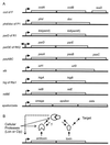
FIG. 1
FIG. 1
(A) Genetic organization of plasmid-encoded TA loci. In all cases but one (the hig locus), the antitoxins are encoded by the upstream gene of the TA operons. Arrows pointing right indicate promoters upstream of the genes. The arrow pointing left indicates a divergent promoter that promotes transcription into the parABC genes of RK2. The resD gene downstream of the ccd genes in F encodes a site-specific resolvase that resolves F multimers into monomers (43, 66). The derivation of gene designations follows: ccd, coupled cell division; phd, prevention of host death; doc, death on curing; kis, killing suppression; kid, killing determinant; pem, plasmid emergency maintenance; pas and stb, plasmid stability; hig, host inhibition of growth; rel, relaxed control of stable RNA synthesis. The pas locus is from the T. ferrooxidans plasmid pTF-CF2; stb is from the S. flexneri plasmid pMYSH6000; ω-ɛ-ζ is from pSM19025 of S. pyogenes; relBE homologues are present on plasmid P307 of E. coli, plasmid pJK2 of A. europaeus, plasmid R485 of M. morganii, and plasmid pRJF2 of B. fibrisolvens. Genes were not drawn to scale. (B) General genetic and functional setup of the TA loci. The antitoxins neutralize the toxins by forming tight complexes with them. The TA complexes bind to operators in the promoter regions and repress transcription (shown by broken arrow pointing to the promoter region). Cellular proteases (Lon or Clp) degrade the antitoxins, thereby leading to activation of the toxins in plasmid-free cells and perhaps during other, as yet unknown, conditions. The question mark indicates that it is not yet known if the antitoxins are degraded when complexed with the toxins or if the toxins and antitoxins dissociate before the antitoxins are degraded.
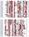
FIG. 2
FIG. 2
Multiple-sequence alignment of 27 RelE proteins from gram-negative and gram-positive bacteria and from archaea. Only RelE proteins with an upstream RelB partner were included in the alignment shown. The characteristics of the corresponding RelB partner proteins are listed in Table 3. Positively charged amino acids are shown in red, and negatively charged amino acids are shown in black. Note the conserved arginine at +82, and the positively charged amino acid at +112 (lysine or arginine). The primary alignment were accomplished by using the Wisconsin GCG package version 8.1.0(a). The multiple-sequence alignment file (msf) was transferred to ClustalX, and the final alignment was edited by eye, using the program Genedoc. The different species and plasmids from which the RelE homologues were derived are indicated by the following letters and numbers after the RelE- suffix: HP1 and HP2, H. pylori homologues 1 and 2, respectively; BF, B. fibrisolvens plasmid pRJF2; SP1, S. pneumoniae homologue 1; AE, A. europaeus plasmid pJK21; SOS, E. coli K-12 homologue 2; HI, H. influenzae; AF3, A. fulgidus homologue 3; St, S. enterica serovar Typhi; pPS, P. shigelloides plasmid; K12, E. coli K-12 homologue 1; MM, M. morganii plasmid R485; P307, E. coli plasmid P307; pB171, E. coli plasmid pB171; VC, V. cholerae; AF1, A. fulgidus homologue 1; MJ1, M. jannaschii homologue 1; Pyr, P. horikoshii OT3; AF2, A. fulgidus homologue 2; BT, B. thuringiensis; MT1 and MT2, M. tuberculosis homologues 1 and 2, respectively; TF, T. ferrooxidans plasmid TF-CF2, AQ, A. aeolicus; AF4, A. fulgidus homologue 4. For simplicity, the irregular RelE homologue of Synechocystis (120 amino acids) was omitted from the alignment. After completion of the database searches, RelE homologues in the unfinished genomes of Salmonella enterica serovars Typhimurium and Paratyphi and Klebsiella pneumoniae were identified. Gaps introduced to maximize alignment are indicated by the dashes.
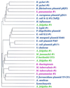
FIG. 3
FIG. 3
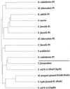
Chladogram (unrooted evolutionary tree) of RelE homologues in prokaryotes. The tree was calculated by PILEUP in the Wisconsin GCG package version 8.1.0(a). The aligned sequences shown in Fig. 2 were used as input. The lengths of horizontal lines indicate relative evolutionary distances, whereas the lengths of the vertical bars are arbitrary. The gram-negative bacterial species are shown in blue, the gram-positive bacterial species are shown in red, and the archaeal species are shown in green. For clarity, the deep-branching organism Aquifex aeolicus was grouped with the gram-negative bacteria.
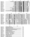
FIG. 4
FIG. 4
Multiple-sequence alignment of 15 ChpK proteins from gram-negative and gram-positive bacteria. Only ChpK proteins with identified putative antitoxin partners were included in the alignment shown. Amino acids with >80% conservation are shown by the black background. Note the fully conserved proline at position 37 and the fully conserved RP motif at positions +47 and +48. The different species and plasmids from which the ChpK (or PemK) homologues were derived are indicated by the following letters and numbers after the ChpK- (or PemK-) suffix: Dr2, D. radiodurans homologue 2; Mt1, M. tuberculosis homologue 1; Bsu1, B. subtilis homologue 1; Sa, S. aureus; Ef and Ef2, E. faecalis homologues 1 and 2, respectively; Mt2, M. tuberculosis homologue 2; Ef3, E. faecalis homologue 3; Pa, P. acidilactici; Dr1, D. radiodurans homologue 1; Tf, T. ferrooxidans; K12, E. coli K-12; R466B, M. morganii plasmid R446B. Gaps introduced to maximize alignment are indicated by the dashes.
FIG. 5
FIG. 5
Chladogram of the ChpK homologues from bacteria. The tree was calculated by PILEUP in the Wisconsin GCG package version 8.1.0(a). The aligned sequences shown in Fig. 4 were used as input. The organisms above the thick line in the figure are gram-positive bacteria, while those below the line are gram-negative bacteria.

Similar articles

Cited by

References

    1. Aizenman E, Engelberg-Kulka H, Glaser G. An Escherichia coli chromosomal “addiction module” regulated by 3′,5′-bispyrophosphate: a model for programmed bacterial cell death. Proc Natl Acad Sci USA. 1996;93:6059–6063. - PMC - PubMed
    1. Altschul S F, Madden T L, Schaffer A A, Zhang J, Zhang Z, Miller W, Lipman D J. Gapped BLAST and PSI-BLAST: a new generation of protein database search programs. Nucleic Acids Res. 1997;25:3389–3402. - PMC - PubMed
    1. Bahassi E M, O'Dea M H, Allali N, Messens J, Gellert M, Couturier M. Interactions of CcdB with DNA gyrase. Inactivation of GyrA, poisoning of the gyrase-DNA complex, and the antidote action of CcdA. J Biol Chem. 1999;274:10936–10944. - PubMed
    1. Baum J. Tn5401, a new class II transposable element from Bacillus thuringiensis. J Bacteriol. 1994;176:2835–2845. - PMC - PubMed
    1. Bech F W, Jørgensen S T, Diderichsen B, Karlström O H. Sequence of the relB transcription unit from Escherichia coli and identification of the relB gene. EMBO J. 1985;4:1059–1066. - PMC - PubMed

Publication types

LinkOut - more resources