

Bacteriophage T4 self-assembly: localization of gp3 and its role in determining tail length
- PMID: 10633101
- PMCID: PMC94330
- DOI: 10.1128/JB.182.3.680-688.2000
Bacteriophage T4 self-assembly: localization of gp3 and its role in determining tail length
Abstract
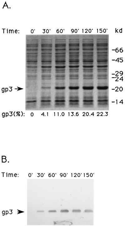
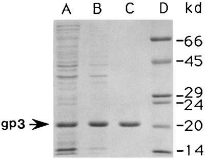
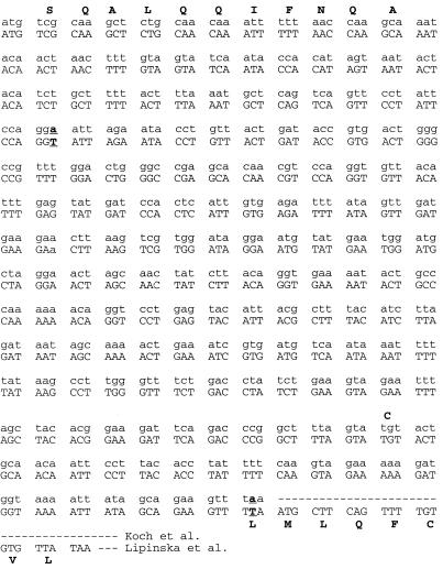
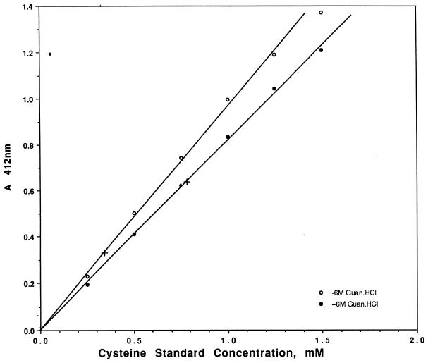
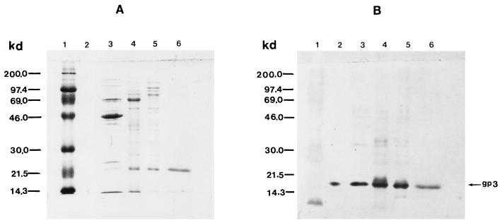
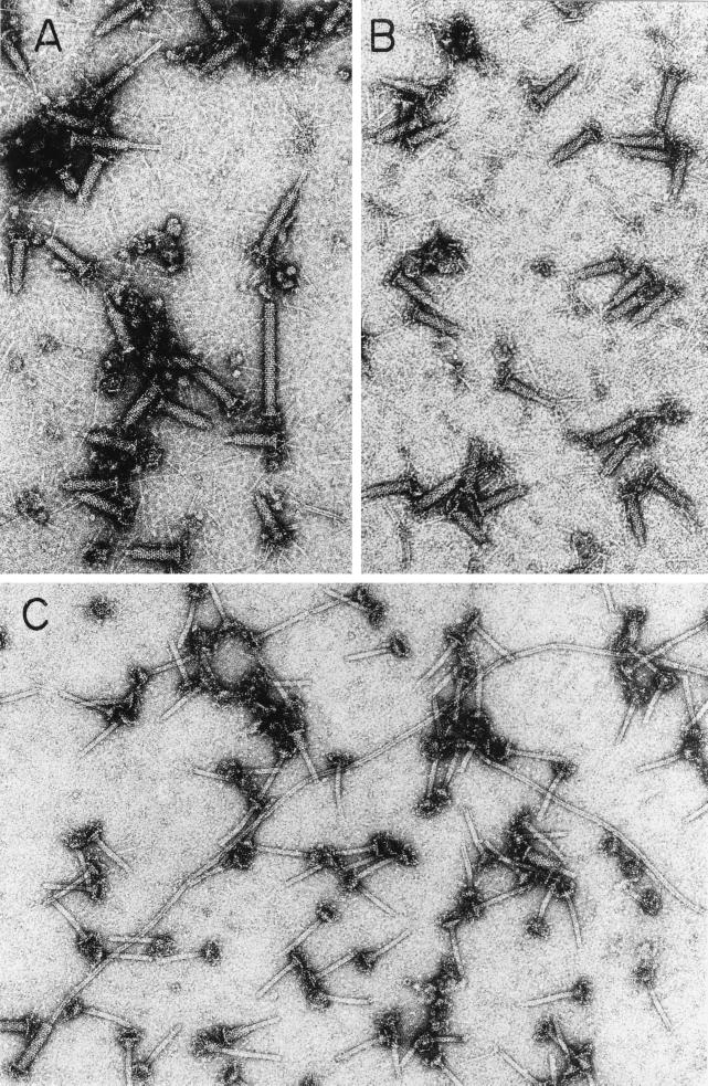
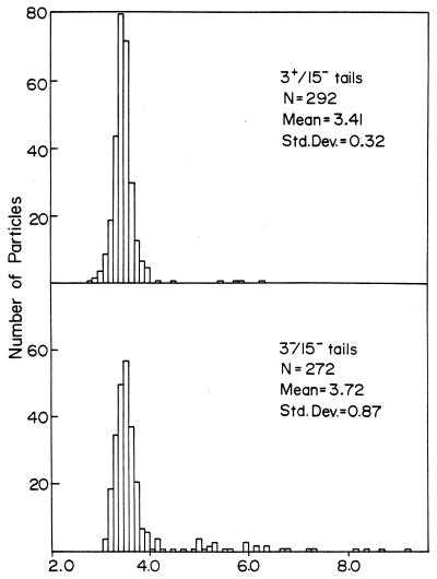
Gene 3 of bacteriophage T4 participates at a late stage in the T4 tail assembly pathway, but the hypothetical protein product, gp3, has never been identified in extracts of infected cells or in any tail assembly intermediate. In order to overcome this difficulty, we expressed gp3 in a high-efficiency plasmid expression vector and subsequently purified it for further analysis. The N-terminal sequence of the purified protein showed that the initial methionine had been removed. Variant C-terminal amino acid sequences were resolved by determining the cysteine content of the protein. The molecular mass of 20.6 kDa for the pure protein was confirmed by Western blotting, using a specific anti-gp3 serum for which the purified protein was the immunogen. We also demonstrated, for the first time, the physical presence of gp3 in the mature T4 phage particle and localized it to the tail tube. By finding a nonleaky, nonpermissive host for a gene 3 mutant, we could clearly demonstrate a new phenotype: the slow, aberrant elongation of the tail tube in the absence of gp3.
Figures

Similar articles
-
P15 and P3, the tail completion proteins of bacteriophage T4, both form hexameric rings.J Bacteriol. 2003 Mar;185(5):1693-700. doi: 10.1128/JB.185.5.1693-1700.2003. J Bacteriol. 2003. PMID: 12591887 Free PMC article.
-
Assembly of the bacteriophage T4 replication machine requires the acidic carboxy terminus of gene 32 protein.J Mol Biol. 1993 Jan 20;229(2):398-418. doi: 10.1006/jmbi.1993.1042. J Mol Biol. 1993. PMID: 8429554
-
Tail length determination in bacteriophage T4.Virology. 1994 Mar;199(2):301-10. doi: 10.1006/viro.1994.1128. Virology. 1994. PMID: 8122363
-
Structure and function of bacteriophage T4.Future Microbiol. 2014;9(12):1319-27. doi: 10.2217/fmb.14.91. Future Microbiol. 2014. PMID: 25517898 Free PMC article. Review.
-
Molecular architecture of bacteriophage T4.Biochemistry (Mosc). 2004 Nov;69(11):1190-202. doi: 10.1007/s10541-005-0064-9. Biochemistry (Mosc). 2004. PMID: 15627372 Review.
Cited by
-
Structural remodeling of bacteriophage T4 and host membranes during infection initiation.Proc Natl Acad Sci U S A. 2015 Sep 1;112(35):E4919-28. doi: 10.1073/pnas.1501064112. Epub 2015 Aug 17. Proc Natl Acad Sci U S A. 2015. PMID: 26283379 Free PMC article.
-
About bacteriophage tail terminator and tail completion proteins: structure of the proximal extremity of siphophage T5 tail.J Virol. 2025 Jan 31;99(1):e0137624. doi: 10.1128/jvi.01376-24. Epub 2024 Dec 23. J Virol. 2025. PMID: 39714170 Free PMC article.
-
The molecular architecture of the bacteriophage T4 neck.J Mol Biol. 2013 May 27;425(10):1731-44. doi: 10.1016/j.jmb.2013.02.012. Epub 2013 Feb 19. J Mol Biol. 2013. PMID: 23434847 Free PMC article.
-
Fine-scale time-lapse analysis of the biphasic, dynamic behaviour of the two Vibrio cholerae chromosomes.Mol Microbiol. 2006 Jun;60(5):1164-78. doi: 10.1111/j.1365-2958.2006.05175.x. Mol Microbiol. 2006. PMID: 16689793 Free PMC article.
-
Structural Studies of the Phage G Tail Demonstrate an Atypical Tail Contraction.Viruses. 2021 Oct 18;13(10):2094. doi: 10.3390/v13102094. Viruses. 2021. PMID: 34696524 Free PMC article.
References
-
- Abuladze N K, Gingery M, Tsai J, Eiserling F A. Tail length determination in bacteriophage T4. Virology. 1994;199:301–310. - PubMed
-
- Ben-Bassat A, Bauer K. Amino-terminal processing of proteins. Nature. 1987;326:315.
-
- Berget P B, King J. T4 tail morphogenesis. In: Mathews C K, Kutter E M, Mosig G, Berget P B, editors. Bacteriophage T4. Washington, D.C.: American Society for Microbiology; 1983. pp. 246–258.
-
- Boyer H W, Roulland-Dussoix D. A complementation analysis of the restriction and modification of DNA in Escherichia coli. J Mol Biol. 1969;41:459–472. - PubMed
-
- Bradford M M. A rapid and sensitive method for the quantitation of microgram quantities of protein utilizing the principle of protein-dye binding. Anal Biochem. 1976;72:248–254. - PubMed
Publication types
MeSH terms
Substances
Grants and funding
LinkOut - more resources
Full Text Sources
Other Literature Sources
