. 2000 Feb;182(3):855-8.
doi: 10.1128/JB.182.3.855-858.2000.

Posttranslational processing of Methanococcus voltae preflagellin by preflagellin peptidases of M. voltae and other methanogens

Affiliations

Posttranslational processing of Methanococcus voltae preflagellin by preflagellin peptidases of M. voltae and other methanogens

J D Correia et al. J Bacteriol. 2000 Feb.

Abstract

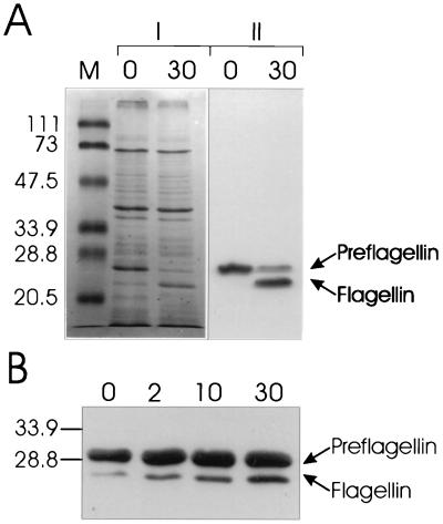
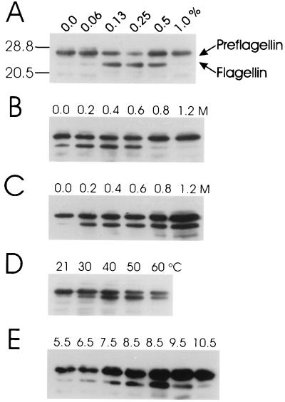
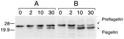
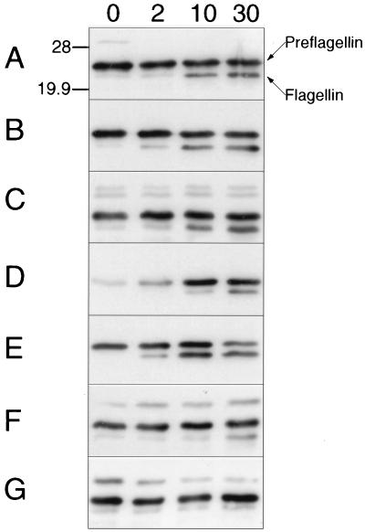
Methanococcus voltae is a mesophilic archaeon with flagella composed of flagellins that are initially made with 11- or 12-amino-acid leader peptides that are cleaved prior to incorporation of the flagellin into the growing filament. Preflagellin peptidase activity was demonstrated in immunoblotting experiments with flagellin antibody to detect unprocessed and processed flagellin subunits. Escherichia coli membranes containing the expressed M. voltae preflagellin (as the substrate) were combined in vitro with methanogen membranes (as the enzyme source). Correct processing of the preflagellin to the mature flagellin was also shown directly by comparison of the N-terminal sequences of the two flagellin species. M. voltae preflagellin peptidase activity was optimal at 37 degrees C and pH 8.5 and in the presence of 0.4 M KCl with 0.25% (vol/vol) Triton X-100.


Figures

FIG. 1
FIG. 1
Processing of M. voltae preflagellins FlaB1 and FlaB2 by the M. voltae preflagellin peptidase. (A) The preflagellin peptidase reaction was performed with approximately 72 μg of induced E. coli KJ91 membranes (FlaB2 substrate source) combined with approximately 18 μg of M. voltae membranes (enzyme source) in 25 mM HEPES buffer (pH 8.5) containing 0.25% (vol/vol) Triton X-100 and 0.4 M KCl (optimized conditions), at a reaction temperature of 37°C. Ten-μl samples were taken at 0- and 30-min time points and immediately mixed with 15 μl of electrophoresis sample buffer and boiled for 5 min. Twenty- and 10-μl samples were analyzed by Coomassie blue staining (|) and immunoblotting with a primary antibody dilution of 1:10,000 (∥), respectively. The relative mobility of prestained SDS-polyacrylamide gel electrophoresis low-range molecular mass standards (Bio-Rad) are indicated in kilodaltons (lane M). (B) The standard preflagellin peptidase reaction was performed with approximately 72 μg of induced E. coli KJ202 membranes (FlaB1 substrate source) combined with approximately 18 μg of M. voltae membranes (enzyme source) in 25 mM HEPES buffer (pH 7.5) containing 0.5% (vol/vol) Triton X-100. Samples removed at 0, 2, 10, and 30 min were similarly prepared and 10-μl aliquots were examined by immunoblotting with anti-FlaB2 antiserum. The positions of 33.9- and 28.8-kDa molecular mass markers are indicated.
FIG. 2
FIG. 2
Optimization of the in vitro preflagellin peptidase reaction for detergent, salt, temperature, and pH. For all reactions, 10-μl samples were removed at 10 min and then immediately mixed with 15 μl of electrophoresis sample buffer and boiled for 5 min; 10-μl aliquots were examined by immunoblotting with a primary antibody dilution of 1:10,000. (A) The standard preflagellin peptidase reaction was performed with approximately 72 μg of induced E. coli KJ91 membranes (substrate source) combined with approximately 18 μg of M. voltae membranes (enzyme source) in 25 mM HEPES buffer (pH 7.5) containing varying final concentrations of Triton X-100 (0.0, 0.0625, 0.125, 0.25, 0.5, and 1.0% [vol/vol]). (B) The standard preflagellin peptidase reaction (25 mM HEPES [pH 7.5] containing 0.5% [vol/vol] Triton X-100) was supplemented with KCl to a final concentration of 0.0, 0.2, 0.4, 0.6, 0.8, or 1.2 M. (C) The standard preflagellin peptidase reaction was supplemented with NaCl to a final concentration of 0.0, 0.2, 0.4, 0.6, 0.8, or 1.2 M. (D) The standard preflagellin peptidase reaction was performed at reaction temperatures of 21, 30, 40, 50, and 60°C. (E) The standard preflagellin peptidase reaction was performed in 25 mM 2-(N-morpholino)ethanesulfonic acid buffer (pH 5.5 and 6.5), 25 mM HEPES buffer (pH 7.5 and 8.5) and 25 mM 1,3,-bis[tris(hydroxymethyl)-methylamino]propane buffer (pH 8.5, 9.5, and 10.5).
FIG. 3
FIG. 3
Comparison of the standard and optimized reaction conditions for the in vitro processing of M. voltae FlaB2 preflagellin by the M. voltae preflagellin peptidase. The standard preflagellin peptidase reaction (A) was performed with approximately 72 μg of induced E. coli KJ91 membranes (substrate source) combined with approximately 18 μg of M. voltae membranes (enzyme source) in 25 mM HEPES buffer (pH 7.5) containing 0.5% (vol/vol) Triton X-100 and the optimized preflagellin peptidase reaction (B) was performed in 25 mM HEPES buffer (pH 8.5) containing 0.4 M KCl and 0.25% (vol/vol) Triton X-100. Ten-microliter samples were removed at 0, 2, 10, and 30 min and then immediately mixed with 15 μl of electrophoresis sample buffer and boiled for 5 min; 10-μl aliquots were examined by immunoblotting with a primary antibody dilution of 1:10,000. The positions of 28- and 19.9-kDa molecular mass markers are indicated.
FIG. 4
FIG. 4
In vitro processing of M. voltae FlaB2 preflagellin by the preflagellin peptidase of M. voltae and other methanococci. The standard preflagellin peptidase reaction was performed with approximately 72 μg of induced E. coli KJ91 membranes (substrate source) combined with approximately 18 μg of M. voltae membranes (A) in 25 mM HEPES buffer (pH 7.5) containing 0.5% (vol/vol) Triton X-100, at a reaction temperature of 37°C. The same reaction was performed with M. deltae membranes (B), M. maripaludis membranes (C), or M. vannielii membranes (D) in place of M. voltae membranes. The same reaction was performed at a reaction temperature of 60°C with M. thermolithotrophicus membranes (E), M. jannaschii membranes (F), or M. igneus membranes (G) as the enzyme source. Ten-microliter samples were removed at 0, 2, 10, and 30 min and then immediately mixed with 15 μl of electrophoresis sample buffer and boiled for 5 min; 10-μl aliquots were examined by immunoblotting with a primary antibody dilution of 1:10,000. Positions of 28- and 19.9-kDa molecular mass markers are indicated.

References

    1. Albers S-V, Konings W N, Driessen A J M. A unique short signal sequence in membrane-anchored proteins of Archaea. Mol Microbiol. 1999;31:1595–1596. - PubMed
    1. Bayley D P, Florian V, Klein A, Jarrell K F. Flagellin genes of Methanococcus vannielii: amplification by the polymerase chain reaction, demonstration of signal peptides and identification of major components of the flagellar filament. Mol Gen Genet. 1998;258:639–645. - PubMed
    1. Bayley D P, Jarrell K F. Overexpression of Methanococcus voltae flagellin subunits in Escherichia coli and Pseudomonas aeruginosa: a source of archaeal preflagellin. J Bacteriol. 1999;181:4146–4153. - PMC - PubMed
    1. Bayley D P, Jarrell K F. Further evidence to suggest that archaeal flagella are related to bacterial type IV pili. J Mol Evol. 1998;46:370–373. - PubMed
    1. Bröckl G, Behr M, Farby S, Hensel R, Kaudewitz H, Biendel E, König H. Analysis and nucleotide sequence of the genes encoding the surface-layer glycoproteins of the hyperthermophilic methanogens Methanothermus fervidus and Methanothermus sociabilis. Eur J Biochem. 1991;199:147–152. - PubMed

Publication types

MeSH terms

LinkOut - more resources