Comparative Study
. 2000 Jan;12(1):165-78.

Osmotic stress induces rapid activation of a salicylic acid-induced protein kinase and a homolog of protein kinase ASK1 in tobacco cells

Affiliations
Comparative Study

Osmotic stress induces rapid activation of a salicylic acid-induced protein kinase and a homolog of protein kinase ASK1 in tobacco cells

M Mikołajczyk et al. Plant Cell. 2000 Jan.

Erratum in

  • Plant Cell 2000 Apr;12(4):611

Abstract

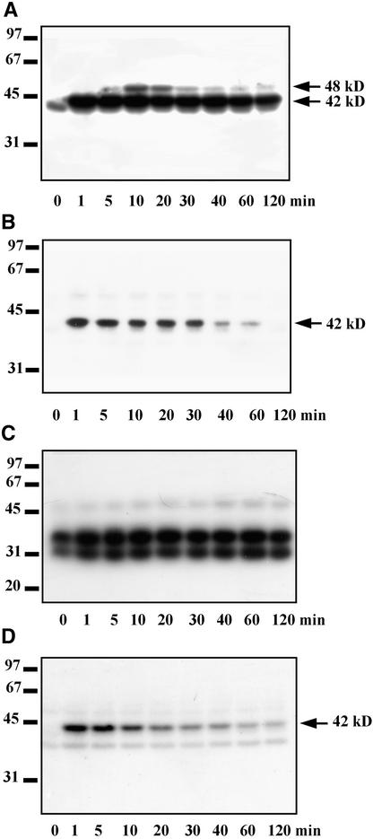
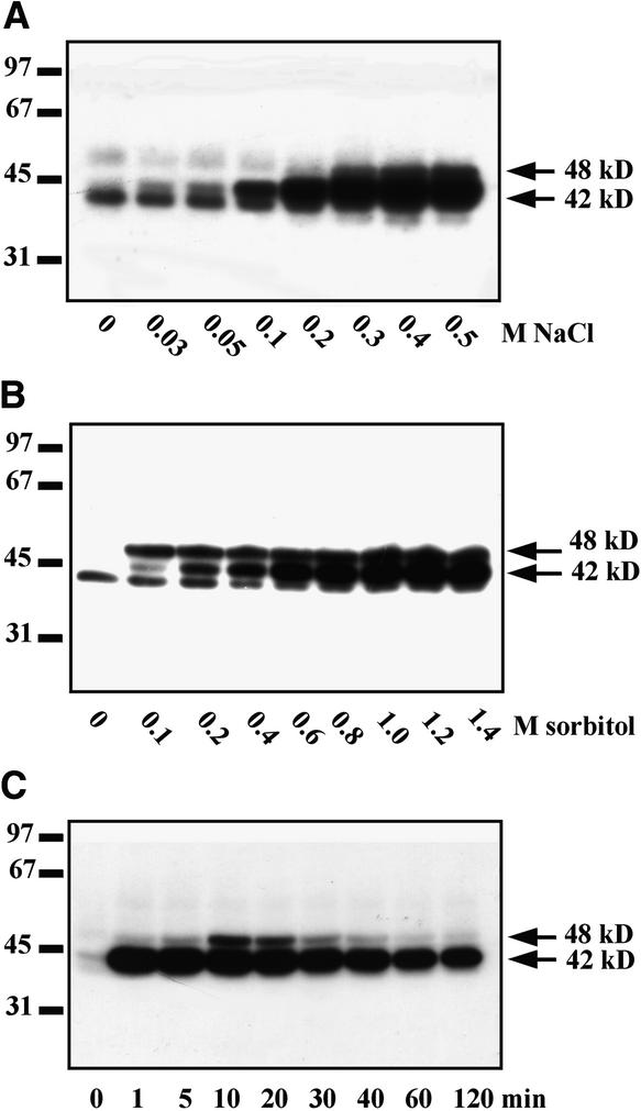
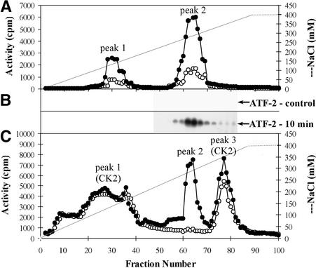
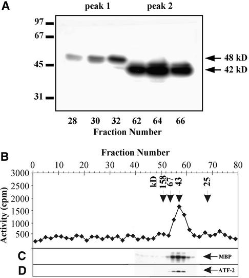
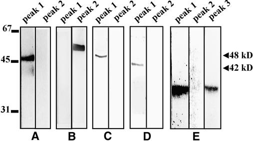
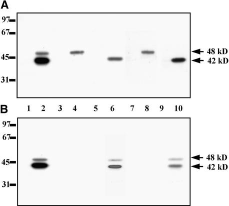
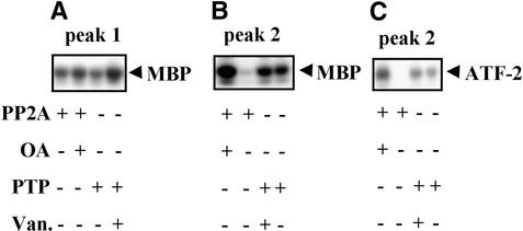
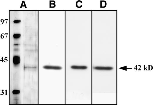
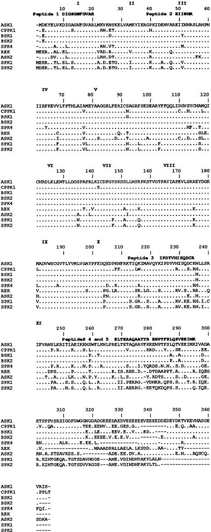
In tobacco cells, osmotic stress induced the rapid activation of two protein kinases that phosphorylate myelin basic protein. Immunological studies demonstrated that the 48-kD kinase is the salicylic acid-induced protein kinase (SIPK), a member of the mitogen-activated protein kinase family. SIPK was activated 5 to 10 min after the cells were exposed to osmotic stresses, and its activity persisted for approximately 30 min. In contrast, the 42-kD kinase was activated within 1 min after osmotic stress, and its activity was maintained for approximately 2 hr. Moreover, in addition to myelin basic protein, the 42-kD kinase phosphorylated casein and two transcription factors, c-Jun and ATF-2. This latter enzyme was inactivated by a serine/threonine-specific phosphatase but, unlike SIPK, was not affected by a tyrosine-specific phosphatase. After the 42-kD kinase was purified to apparent homogeneity, tryptic peptide analysis indicated that it is a homolog of Arabidopsis serine/threonine kinase1 (ASK1).


Figures

Figure 1.
Figure 1.
Identification of Protein Kinases Activated by Salinity Stress. Tobacco suspension culture cells were treated with 250 mM NaCl. Aliquots of the culture were taken at the indicated times, and kinase activities were analyzed in cell cytosolic extracts by the in-gel kinase assays using MBP, recombinant GST–ATF-2, or casein as the substrates. Molecular mass markers are given at left in kilodaltons. The 42 and 48 kD at right indicate molecular masses of the protein kinases activated by salinity stress. (A) Identification of protein kinases that phosphorylate MBP. (B) Identification of protein kinases that phosphorylate ATF-2. (C) and (D) Identification of protein kinases that phosphorylate casein. The assay shown in (D) was performed in the presence of 100 μg/mL heparin.
Figure 2.
Figure 2.
Activation of Protein Kinases in Tobacco Cells Exposed to NaCl and Sorbitol Stress. (A) and (B) Dose–response. Tobacco cells were treated with various concentrations of NaCl (A) or sorbitol (B) for 10 min, and protein kinase activities were monitored in cell extracts by using an in-gel kinase assay with MBP as substrate. (C) Time course. Tobacco cells were treated with 900 mM sorbitol. Aliquots of the culture were taken at the indicated times, and kinase activities were analyzed in cell extracts by the in-gel kinase assay with MBP as substrate. Molecular mass markers are given at left in kilodaltons. The 42 and 48 kD at right indicate molecular masses of the protein kinases activated by salinity stress.
Figure 3.
Figure 3.
Elution Profile of Protein Kinases from the Mono Q HR 5/5 Column. Chromatography of cytosolic extracts of tobacco cells treated for 10 min with 250 mM NaCl (filled circles) and untreated (control) cells (open circles) on a Mono Q column was performed. Protein kinase activities were determined by an in-solution kinase assay (see Methods). (A) Analysis of protein kinase activities that phosphorylate MBP. (B) Analysis of protein kinase activities that phosphorylate ATF-2. (C) Analysis of protein kinase activities that phosphorylate casein.
Figure 4.
Figure 4.
Estimation of Molecular Masses of Protein Kinases Activated by Osmotic Stress. (A) Enzymes eluted from the Mono Q column in peak 1 (fractions 28, 30, and 32) and peak 2 (fractions 62, 64, and 66) were analyzed by using an in-gel kinase assay with the MBP. Molecular mass markers are indicated in kilodaltons at left. The 42 and 48 kD at right indicate molecular masses of the protein kinases activated by salinity stress. (B) to (D) The protein kinases eluted in peak 2 were chromatographed on the Superose 12 column. Protein kinase activities were assayed by using casein (B), MBP (C), or ATF-2 (D) as substrate. Arrowheads and numbers in kilodaltons at top indicate molecular mass markers.
Figure 5.
Figure 5.
Immunological Analysis of Tobacco Protein Kinases Activated by Osmotic Stress. For protein gel blot analysis of proteins eluted from the Mono Q column in peaks 1 and 2 and occasionally in peak 3, the blots were probed with the following antibodies: (A) Rabbit polyclonal anti–ERK1/ERK2 antibodies. (B) Rabbit polyclonal anti-JNK1 antibodies. (C) Rabbit polyclonal anti-SIPK antibodies. (D) Rabbit polyclonal anti-WIPK antibodies. (E) Rabbit polyclonal anti-CK2α antibodies. Molecular mass markers are given in kilodaltons at left; 42 and 48 kD at right indicate molecular masses of the protein kinases activated by salinity stress.
Figure 6.
Figure 6.
SIPK-Specific Antibodies Immunoprecipitate the 48-kD Osmotic Stress–Activated Protein Kinase. Protein extracts (50 μg) from untreated cells (lanes 1, 3, 5, 7, and 9), from cells exposed to 900 mM sorbitol for 10 min (lanes 2, 4, and 6), or to 250 mM NaCl (lanes 8 and 10) were analyzed by using an in-gel kinase assay with MBP as substrate, as described in Methods, before and after immunoprecipitation with anti-SIPK or anti-WIPK antibodies. (A) Immunoprecipitation with anti-SIPK antibodies. (B) Immunoprecipitation with anti-WIPK antibodies. Lanes 1 and 2 show kinase activity of protein extracts not incubated with antibodies; lanes 3, 4, 7, and 8 show kinase activity of immunocomplexes; and lanes 5, 6, 9, and 10 show proteins possessing kinase activity that were not bound to antibodies, remaining in the supernatant after immunoprecipitation. Molecular mass markers are given in kilodaltons at left; 42 and 48 kD indicate molecular masses of the osmotic stress–activated protein kinases.
Figure 7.
Figure 7.
Inhibition by Treatment with Protein Phosphatases of the Protein Kinases Activated by Osmotic Stress. Kinases eluted in peaks 1 and 2 from the Mono Q column were preincubated for 20 min with PP2A or PTP at 30°C in the presence (controls) or absence of specific phosphatase inhibitors ocadaic acid (OA) for PP2A and sodium vanadate (Van.) for PTP. (+) and (−) indicate the presence and absence, respectively, of protein phosphatase or inhibitor in the reaction mixture. (A) Effect of dephosphorylation on activities of protein kinase(s) eluted in peak 1 measured with MBP as the substrate. (B) Effect of dephosphorylation on activities of protein kinase(s) eluted in peak 2 measured with MBP as the substrate. (C) Effect of dephosphorylation on activities of protein kinase(s) eluted in peak 2 measured with ATF-2 as the substrate. Phosphorylation of MBP and ATF-2 was determined by autoradiography of phosphorylated substrates resolved by SDS-PAGE.
Figure 8.
Figure 8.
Elution Profiles of Kinase Activity and Protein from Each Purification Step. (A) Chromatography on a SOURCE 15Q column. (B) Chromatography on a phenyl-Sepharose column. (C) Chromatography on a heparin-Sepharose column. (D) Chromatography on a Mono Q column. Kinase activity (filled circles) was determined by the in-solution kinase assay with casein as substrate. For inhibition of casein kinases, heparin was added to the incubation mixtures at a concentration of 100 μg/mL. The dashed lines indicate NaCl gradients, and lines with open diamonds indicate protein concentration.
Figure 9.
Figure 9.
The 42-kD Osmotic Stress–Activated Protein Kinase Phosphorylates MBP and Casein Equally Well. Substrate specificity of the purified 42-kD osmotic stress–activated protein kinase was analyzed by using an in-gel kinase assay. (A) SDS-PAGE of the purified 42-kD osmotic stress–activated protein kinase. Proteins were visualized by silver staining. (B) In-gel kinase assay of the 42-kD protein kinase with MBP. (C) In-gel kinase assay of the 42-kD protein kinase with casein. (D) In-gel kinase assay of the 42-kD protein kinase with equal weights of MBP and casein.
Figure 10.
Figure 10.
Comparison of Tryptic Peptide Sequences of the 42-kD Kinase with Other Proteins. Proteins exhibiting the greatest sequence similarities to peptides derived from the 42-kD kinase are shown. Dots represent amino acid residues that are identical to ASK1 (Park et al., 1993). References for the sequences of the other kinases are as follows: CPPK1 (P. Heino, M. Nylander, T. Palva, and D. Bartels, unpublished results; GenBank accession number CAA06503), BSK1 (Park et al., 1995), BSK2 (Park et al., 1995), SPK4 (Yoon et al., 1997), rice endosperm protein kinase (REK; Hotta et al., 1998), ASK2 (Park et al., 1993), soybean protein kinase 1 (SPK1; P.G. Shin, H. Yoon, Y.H. Jeong, J. Bahk, J.C. Hong, and M.J. Cho, unpublished results; GenBank accession number AAA33979), and soybean protein kinase 2 (SPK2; P.G. Shin, H. Yoon, M. Kim, J. Bahk, J.C. Hong, and M.J. Cho, unpublished results; GenBank accession number AAA34017). The conserved subdomains of the serine/threonine kinase domain are indicated by roman numerals. Gaps are marked with dashes and were introduced to maximize alignment. The peptide sequences obtained by microsequencing are shown above the ASK1 sequence.

References

    1. Adam, A.L., Pike, S., Hoyos, E., Stone, J.M., Walker, J.C., and Novacky, A. (1997). Rapid and transient activation of a myelin basic protein kinase in tobacco leaves treated with harpin from Erwinia amylovora. Plant Physiol. 115 853–861. - PMC - PubMed
    1. Anderberg, R.J., and Walker-Simmons, M.K. (1992). Isolation of a wheat cDNA clone for an abscisic acid–inducible transcript with homology to protein kinases. Proc. Natl. Acad. Sci. USA 89 10183–10187. - PMC - PubMed
    1. Bögre, L., Ligterink, W., Heberle-Bors, E., and Hirt, H. (1996). Mechanosensors in plants. Nature 383 489–490. - PubMed
    1. Bögre, L., Ligterink, W., Meskiene, I., Barker, P.J., Heberle-Bors, E., Huskinsson, N.S., and Hirt, H. (1997). Wounding induces the rapid and transient activation of a specific MAP kinase pathway. Plant Cell 9 75–83. - PMC - PubMed
    1. Bohnert, H.J., Nelson, D.E., and Jensen, R.G. (1995). Adaptation to environmental stresses. Plant Cell 7 1099–1111. - PMC - PubMed

Publication types

MeSH terms

Substances

LinkOut - more resources