. 2000 Jan 17;19(2):234-40.
doi: 10.1093/emboj/19.2.234.

A membrane-embedded glutamate is required for ligand binding to the multidrug transporter EmrE

Affiliations

A membrane-embedded glutamate is required for ligand binding to the multidrug transporter EmrE

T R Muth et al. EMBO J. .

Abstract

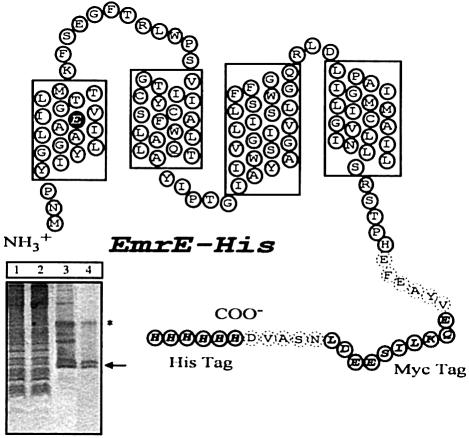
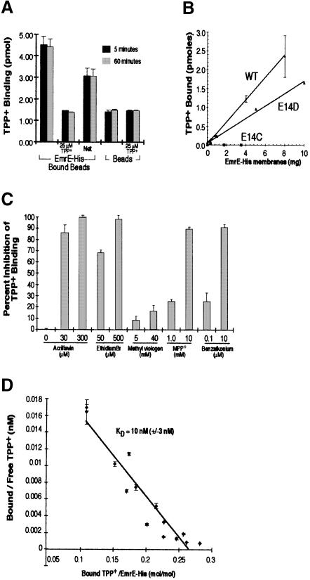
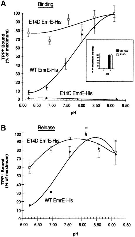
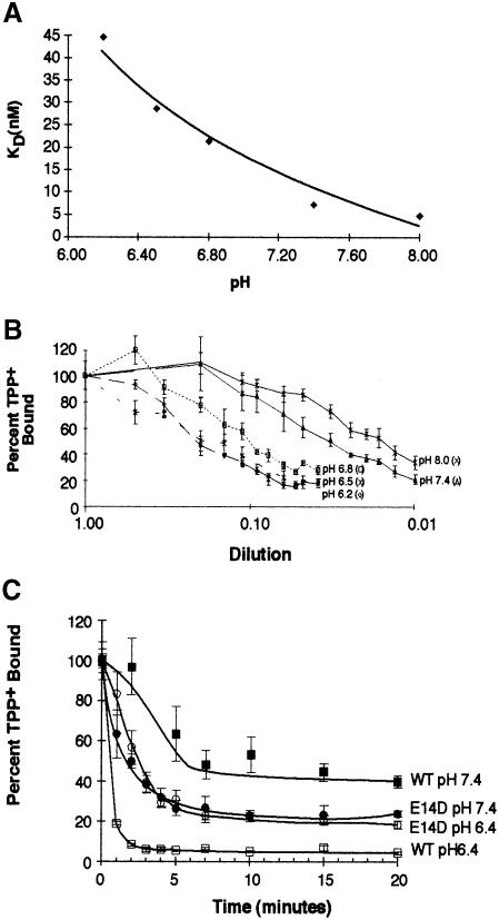
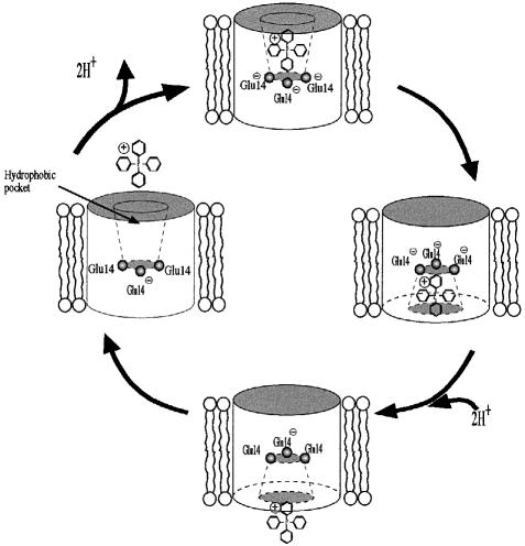
EmrE is an Escherichia coli multidrug transporter that confers resistance to a variety of toxins by removing them in exchange for hydrogen ions. The detergent-solubilized protein binds tetraphenylphosphonium (TPP(+)) with a K(D) of 10 nM. One mole of ligand is bound per approximately 3 mol of EmrE, suggesting that there is one binding site per trimer. The steep pH dependence of binding suggests that one or more residues, with an apparent pK of approximately 7.5, release protons prior to ligand binding. A conservative Asp replacement (E14D) at position 14 of the only membrane-embedded charged residue shows little transport activity, but binds TPP(+) at levels similar to those of the wild-type protein. The apparent pK of the Asp shifts to <5.0. The data are consistent with a mechanism requiring Glu14 for both substrate and proton recognition. We propose a model in which two of the three Glu14s in the postulated trimeric EmrE homooligomer deprotonate upon ligand binding. The ligand is released on the other face of the membrane after binding of protons to Glu14.


Figures

None
Fig. 1. Model of EmrE–His with the four transmembrane regions predicted by hydropathy plots. The His and Myc tags are indicated by open ovals with one-letter amino acid codes shown in bold italicized text. Dashed ovals indicate residues that were incorporated into the transporter in the process of linking in the epitope tags. The Glu14 residue in the first transmembrane region is highlighted. The Myc tag is used here only as a linker to keep the His residues away from the membrane. Without the linker, the His-tagged transporter displays only residual activity. Inset, SDS–PAGE analysis of different stages of purification. Lane 1, total membranes; lane 2, detergent-solubilized extract after Ni–NTA purification, unbound fraction; lane 3, EmrE after Ni–NTA purification; lane 4, EmrE after size exclusion purification. The arrow indicates the monomeric form of EmrE; the asterisk the dimeric form. The apparent Mr of monomeric EmrE–His is 14 400 Da.
None
Fig. 2. Purified EmrE–His specifically binds [3H]TPP+. (A) Purified EmrE–His (4 μg) was bound to Ni–NTA beads and then incubated with 25 nM [3H]TPP+ in NH4–DM buffer. TPP+ binding reactions were incubated for 5 min (black bars) or 60 min (gray bars). Identical binding reactions were performed with beads that had not been bound with EmrE–His. Each TPP+ binding reaction was performed in the presence or absence of 25 μM cold TPP+. (B) Bacterial membranes expressing EmrE–His wild type, E14D or E14C were detergent solubilized and then incubated with Ni–NTA beads. Increasing amounts of solubilized membranes were bound to 20 μl of beads. The beads were then washed to remove proteins that did not bind. The bound beads were then incubated for 30 min with 25 nM [3H]TPP+ and the amount of bound TPP+ was determined as described above. (C) EmrE–beads were incubated with 50 nM [3H]TPP+ in the presence of various EmrE substrates. The concentration of inhibitor used is indicated below each bar. The values of TPP+ binding are plotted in terms of percentage inhibition, with binding in the absence of inhibitors representing zero inhibition. (D) EmrE–His transporter (0.35 μg) bound to Ni–NTA beads was incubated with TPP+ over a range of TPP+ concentrations (10–300 nM). Bound TPP+ was determined by measuring the EmrE–His-associated radioactivity after isolating the beads from the supernatant, and free TPP+ concentrations were measured by subtracting bound radioactivity from the total radioactivity present in each binding reaction. The KD was determined from the reciprocal of the slope obtained from the Scatchard plot. The KD for TPP+ under these conditions was 10 nM (±3 nM). In (A–D), all results are generated from the average of triplicate reactions and the error bars represent the average deviation.
None
Fig. 3. Effect of pH on the binding and release of TPP+ to EmrE–His wild type and the E14 replacement mutants. (A) EmrE–beads were incubated with 25 nM [3H]TPP+ for 1 h in solutions over a range of pH values. Values are graphed as a percentage of maximal TPP+ binding. Purified wild-type EmrE–His (•) and E14D EmrE–His mutant (□) or E14C EmrE–His membranes (▴) were assayed for TPP+ binding over a range of pH values. The inset graph shows TPP+ binding at pH 5 and 8. For this experiment, TPP+ bound to EmrE–His was separated from free TPP+ using size exclusion chromatography over a Sephadex G–50 column. After the TPP+ binding to EmrE–His at the desired pH, the binding reaction was run over the column and the bound [3H]TPP+ collected in scintillation vials by centrifugation and then counted. The black bars represent the data from wild-type EmrE–His and the open bars represent data from the E14D EmrE–His mutant construct. (B) EmrE–beads were incubated with 25 nM TPP+ for 30 min at 4°C. EmrE–beads bound to TPP+ at equilibrium levels were diluted 1:75 in 60 mM buffered solutions at the desired pH and incubated at 4°C for 2 h. This time was determined to be sufficient for binding reactions to reach equilibrium in experiments not shown here. Values are graphed as a percentage of maximal TPP+ binding. Wild-type EmrE–His (•) and the EmrE–His E14D mutant (□) were assayed. For (A) and (B), each point represents the average of triplicate binding reactions. The error bars represent the average deviation of the triplicate measurements.
None
Fig. 4. Effect of pH on the KD of TPP+ binding to EmrE–His and rate of release. EmrE–beads were incubated with 25 nM [3H]TPP+ for 30 min at 4°C. The EmrE–His bound to TPP+ at equilibrium was transferred into release buffer over a range of increasing dilutions and incubated at 4°C for 2 h. EmrE–His bound to TPP+ was separated from the binding solution by pelleting the beads and removing the supernatant. A fraction of the supernatant was counted to determine the concentration of free TPP+. EmrE–His was released from the beads by incubating with 150 mM imidazole buffer and the resulting supernatants were counted to measure the amount of bound TPP+ present. Using Scatchard analysis, we determined a KD for TPP+ binding at several pH values. (A) The resulting KDs were plotted against pH. (B) The percentage of bound TPP+ at each dilution. Each set of points represents the dilution-dependent release at a different pH. The values are plotted as a percentage of maximal TPP+ binding. Each point represents the average of triplicate binding reactions. The error bars represent the average deviation of the triplicate measurements. (C) EmrE–beads were incubated with 25 nM [3H]TPP+ for 30 min at 4°C. EmrE–His bound to TPP+ at equilibrium was diluted in 60 mM buffered solutions at pH 7.4 (▪, •) or pH 6.4 (□, ○) and incubated at 4°C for the times indicated. Values are plotted as a percentage of maximal TPP+ binding. Wild-type EmrE–His (squares) and the E14D EmrE–His (circles) were assayed. Each point represents the average of triplicate binding reactions. The error bars represent the average deviation of the triplicate measurements.
None
Fig. 5. Proposed mechanism of transport by EmrE. This schematic depicts a possible catalytic cycle for TPP+/H+ binding and release from the EmrE transporter. As the substrate approaches the hydrophobic binding pocket, two protons are released from the negatively charged glutamate triplet. The positively charged substrate is bound through electrostatic interactions with the negatively charged carboxylate groups extending from each glutamate residue. Following an unknown conformational transition, the opening to the binding pockets becomes accessible to the alternative face of the membrane, while being closed off from the opposite face. The subsequent movement of two protons towards the binding pocket catalyzes the release of the bound substrate. The transporter then relaxes, undergoing a conformational transition that converts the binding pocket accessibility back to the original membrane face.

References

    1. Arkin I., Russ, W., Lebendiker, M. and Schuldiner, S. (1996) Determining the secondary structure and orientation of EmrE, a multi-drug transporter, indicates a transmembrane four helix bundle. Biochemistry, 35, 7233–7238. - PubMed
    1. Barker E.L., Moore, K.R., Rakhshan, F. and Blakely, R.D. (1999) Transmembrane domain I contributes to the permeation pathway for serotonin and ions in the serotonin transporter. J. Neurosci., 19, 4705–4717. - PMC - PubMed
    1. Edgar R. and Bibi, E. (1999) A single membrane-embedded negative charge is critical for recognizing positively charged drugs by the Escherichia coli multidrug resistance protein MdfA. EMBO J., 18, 822–832. - PMC - PubMed
    1. Gottesman M.M., Pastan, I. and Ambudkar, S.V. (1996) P-glycoprotein and multidrug resistance. Curr. Opin. Genet. Dev., 6, 610–617. - PubMed
    1. Grinius L. and Goldberg, E. (1994) Bacterial multidrug resistance is due to a single membrane protein which functions as a drug pump. J. Biol. Chem., 269, 29998–30004. - PubMed

Publication types

MeSH terms

LinkOut - more resources