. 2000 Jan 17;191(2):313-20.
doi: 10.1084/jem.191.2.313.

Development of chronic inflammatory arthropathy resembling rheumatoid arthritis in interleukin 1 receptor antagonist-deficient mice

Affiliations

Development of chronic inflammatory arthropathy resembling rheumatoid arthritis in interleukin 1 receptor antagonist-deficient mice

R Horai et al. J Exp Med. .

Abstract

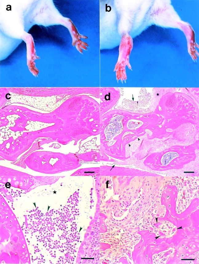
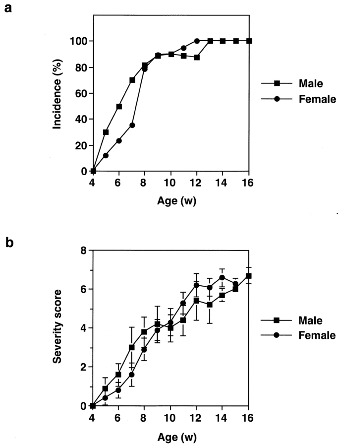
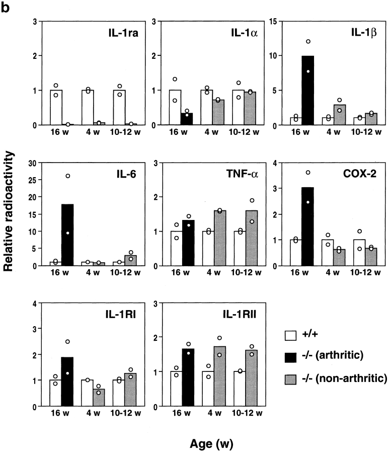
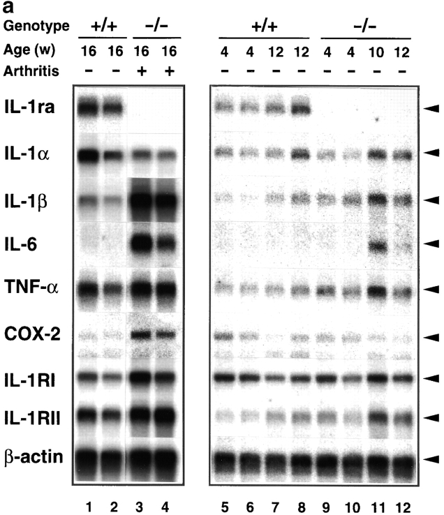
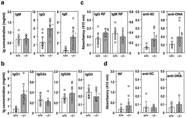
Interleukin (IL)-1 is a proinflammatory cytokine that plays important roles in inflammation, host defense, and the neuro-immuno-endocrine network. IL-1 receptor antagonist (ra) is an endogenous inhibitor of IL-1 and is supposed to regulate IL-1 activity. However, its pathophysiological roles in a body remain largely unknown. To elucidate the roles of IL-1ra, IL-1ra-deficient mice were produced by gene targeting, and pathology was analyzed on different genetic backgrounds. We found that all of the mice on a BALB/cA background, but not those on a C57BL/6J background, spontaneously developed chronic inflammatory polyarthropathy. Histopathology showed marked synovial and periarticular inflammation, with articular erosion caused by invasion of granulation tissues closely resembling that of rheumatoid arthritis in humans. Moreover, elevated levels of antibodies against immunoglobulins, type II collagen, and double-stranded DNA were detected in these mice, suggesting development of autoimmunity. Proinflammatory cytokines such as IL-1beta, IL-6, and tumor necrosis factor alpha were overexpressed in the joints, indicating regulatory roles of IL-1ra in the cytokine network. We thus show that IL-1ra gene deficiency causes autoimmunity and joint-specific inflammation and suggest that IL-1ra is important in maintaining homeostasis of the immune system. Possible involvement of IL-1ra gene deficiency in RA will be discussed.


Figures

Figure 1
Figure 1
Histopathology of the ankle joints of BALB/cA IL-1ra−/− mice. The ankles of a normal IL-1ra+/+ mouse (a) and an affected IL-1ra−/− mouse (b) at 16 wk of age. Swelling and redness of the joints was observed in the IL-1ra−/− mouse. Microscopic observation of the joints of an IL-1ra+/+ mouse (c) and an IL-1ra−/− mouse (d), showing erosive destruction of the bone in the IL-1ra−/− mouse (arrowheads). Infiltration of inflammatory cells (arrows) and proliferation of the lining cells of the synovial membrane (★) were remarkable. (e) Neutrophil infiltration in the articular cavity (arrowheads) and deposition of fibrin clots (★). (f) Pannus formation; note the activation of osteoclasts (arrowheads). Scale bars: c and d, 100 μm; e and f, 50 μm.
Figure 2
Figure 2
Incidence of arthritis in BALB/cA IL-1ra−/− mice. (a) Incidence of arthritis in IL-1ra−/− mice. Development of arthritis was inspected weekly, and the percentage of arthritic mice (grade ≥1) is shown. (b) Arthritic severity score of IL-1ra−/− mice. The average scores of the mice with or without lesions are shown. Male mice: ▪, n = 10; female mice: •, n = 20. Average ± SEM is indicated. w, wk.
Figure 4
Figure 4
Augmentation of IL-1β, IL-6, TNF-α, COX-2, and IL-1R mRNA expression in the joints of BALB/cA IL-1ra−/− mice. Total RNA was isolated from the joints, and poly (A)+ RNA was purified. Expression of the genes for IL-1ra, IL-1α, IL-1β, IL-6, TNF-α, IL-1RI, IL-1RII, and COX-2 was examined by Northern blot hybridization analysis. (a) Lanes 1 and 2 and 5–8, IL-1ra+/+ mice; lanes 3 and 4 and 9–12, IL-1ra−/− mice. Lanes 1 and 2 and 5–12, nonarthritic mice; lanes 3 and 4, arthritic mice (severity score = 6). Ages of the mice are indicated at top. The results were reproducible in three independent experiments. (b) Densitometric analysis of mRNA expression levels. The radioactivity for a band shown in panel a was measured by a BAS 2000 system and normalized by that of β-actin; the radioactivities of the mutant mice relative to those of age-matched wild-type mice are shown. White bar, wild-type mouse; black bar, arthritic IL-1ra−/− mouse; gray bar, nonarthritic IL-1ra−/− mouse. w, wk.
Figure 4
Figure 4
Augmentation of IL-1β, IL-6, TNF-α, COX-2, and IL-1R mRNA expression in the joints of BALB/cA IL-1ra−/− mice. Total RNA was isolated from the joints, and poly (A)+ RNA was purified. Expression of the genes for IL-1ra, IL-1α, IL-1β, IL-6, TNF-α, IL-1RI, IL-1RII, and COX-2 was examined by Northern blot hybridization analysis. (a) Lanes 1 and 2 and 5–8, IL-1ra+/+ mice; lanes 3 and 4 and 9–12, IL-1ra−/− mice. Lanes 1 and 2 and 5–12, nonarthritic mice; lanes 3 and 4, arthritic mice (severity score = 6). Ages of the mice are indicated at top. The results were reproducible in three independent experiments. (b) Densitometric analysis of mRNA expression levels. The radioactivity for a band shown in panel a was measured by a BAS 2000 system and normalized by that of β-actin; the radioactivities of the mutant mice relative to those of age-matched wild-type mice are shown. White bar, wild-type mouse; black bar, arthritic IL-1ra−/− mouse; gray bar, nonarthritic IL-1ra−/− mouse. w, wk.
Figure 3
Figure 3
Antibody production in IL-1ra−/− mice. Serum antibody levels in IL-1ra−/− mice (−/−) and their wild-type littermates (+/+) were determined by ELISA at 16 wk of age. (a) Levels of total IgM, IgG, and IgE in BALB/cA background mice. (b) Levels of IgG subclasses in BALB/cA background mice. (c) Levels of autoantibodies against IgG (RF) of IgG class and IgM class, IIC, and dsDNA in BALB/cA background mice. (d) Autoantibody levels in C57BL/6J background mice. The serum dilution in c and d was 1/25. Autoantibody levels were expressed as the relative absorbance of the ELISA. Average ± SD of each genotype is shown. Open bars: wild-type mice; n = 13 (a), n = 11 (b), n = 9 (c), and n = 8 (d). Shaded bars: IL-1ra−/− mice; n = 14 (a), n = 12 (b), n = 10 (c), and n = 15 (d). Statistical significance was calculated by Student's t test. *P < 0.0001; P < 0.005; § P < 0.05.

References

    1. Firestein, G.S., and N.J. Zvaifler. 1992. Rheumatoid arthritis: a disease of disordered immunity. In Inflammation: Basic Principles and Clinical Correlates, 2nd ed. Raven Press, Ltd., New York. 959–975.
    1. Feldmann M., Brennan F.M., Maini R.N. Role of cytokines in rheumatoid arthritis. Annu. Rev. Immunol. 1996;14:397–440. - PubMed
    1. Pettipher E.R., Higgs G.A., Henderson B. Interleukin 1 induces leukocyte infiltration and cartilage proteoglycan degradation in the synovial joint. Proc. Natl. Acad. Sci. USA. 1986;83:8749–8753. - PMC - PubMed
    1. Marinova-Mutafchieva L., Williams R.O., Mason L.J., Mauri C., Feldmann M., Maini R.N. Dynamics of proinflammatory cytokine expression in the joints of mice with collagen-induced arthritis (CIA) Clin. Exp. Immunol. 1997;107:507–512. - PubMed
    1. van den Berg W.B., Joosten L.A., Helsen M., van de Loo F.A. Amelioration of established murine collagen-induced arthritis with anti-IL-1 treatment. Clin. Exp. Immunol. 1994;95:237–243. - PMC - PubMed

Publication types

MeSH terms