. 2000 Jan 17;191(2):321-34.
doi: 10.1084/jem.191.2.321.

Activation of the p38 mitogen-activated protein kinase pathway arrests cell cycle progression and differentiation of immature thymocytes in vivo

Affiliations

Activation of the p38 mitogen-activated protein kinase pathway arrests cell cycle progression and differentiation of immature thymocytes in vivo

N L Diehl et al. J Exp Med. .

Abstract

The development of T cells in the thymus is coordinated by cell-specific gene expression programs that involve multiple transcription factors and signaling pathways. Here, we show that the p38 mitogen-activated protein (MAP) kinase signaling pathway is strictly regulated during the differentiation of CD4(-)CD8(-) thymocytes. Persistent activation of p38 MAP kinase blocks fetal thymocyte development at the CD25(+)CD44(-) stage in vivo, and results in the lack of T cells in the peripheral immune system of adult mice. Inactivation of p38 MAP kinase is required for further differentiation of these cells into CD4(+)CD8(+) thymocytes. The arrest of cell cycle in mitosis is partially responsible for the blockade of differentiation. Therefore, the p38 MAP kinase pathway is a critical regulatory element of differentiation and proliferation during the early stages of in vivo thymocyte development.


Figures
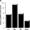
Figure 1
Figure 1
Progressive enlargement of the thymus in the MKK6(Glu) transgenic mice. (A) p38 MAP kinase activity in thymocyte populations from wild-type mice. Whole cell extracts (4 × 10⁵) of freshly isolated total thymocytes (T.T), DN, DP, and mature single CD4+ thymocytes from wild-type mice were assayed for p38 MAP kinase activity using the substrate GST-ATF2. (B) Expression of MKK6(Glu) in the MKK6(Glu) transgenic mice. The expression of endogenous p38 MAP kinase and the MKK6(Glu) transgene (MKK6) was analyzed in thymocytes and spleen cells from negative littermate control mice (NLC) and mice from three different MKK6(Glu) transgenic (Tg+) lines (lines 45, 3, and 6) by Western blot analysis. Blots were probed with either an anti-Flag antibody (MKK6) or an anti-p38 MAP kinase polyclonal antibody (p38). (C) Activation of p38 MAP kinase by the expression of MKK6(Glu) in thymocytes. Total thymocytes (5 × 10⁵ cells) from MKK6(Glu) transgenic mice and negative littermate control mice were lysed, and whole extracts were assayed for p38 activity using the substrate GST-ATF2 or for JNK activity using the substrate GST-c-Jun. (D) Diminished thymocyte number in the MKK6(Glu) transgenic mice. Data represent the percentage of the total number of cells in the thymus from the MKK6(Glu) transgenic mice from different lines compared with the total number of thymocytes in negative littermate control mice. (E) Enlarged thymus in the MKK6(Glu) transgenic mice. Thymi from 3- and 5.5-mo-old (3 m and 5 m, respectively) negative littermate control and MKK6(Glu) transgenic (line 6) mice (left two panels). Thymus (thick arrow) and lungs (thin arrow) from 5.5-mo-old (5 m) negative littermate control and MKK6(Glu) transgenic mice (right two panels). (F) Weight of thymi from 2.5–3.5- and 4.5–5.5-mo-old (3 m and 5 m, respectively) negative littermate control and MKK6(Glu) transgenic mice. Values represent average weight (n = 4). (G) Progressive accumulation of thymocytes in the MKK6(Glu) transgenic mice. Values represent the average (n = 4) of the number of total thymocytes in 3- and 5-mo-old negative littermate control and MKK6(Glu) transgenic mice.
Figure 1
Figure 1
Progressive enlargement of the thymus in the MKK6(Glu) transgenic mice. (A) p38 MAP kinase activity in thymocyte populations from wild-type mice. Whole cell extracts (4 × 10⁵) of freshly isolated total thymocytes (T.T), DN, DP, and mature single CD4+ thymocytes from wild-type mice were assayed for p38 MAP kinase activity using the substrate GST-ATF2. (B) Expression of MKK6(Glu) in the MKK6(Glu) transgenic mice. The expression of endogenous p38 MAP kinase and the MKK6(Glu) transgene (MKK6) was analyzed in thymocytes and spleen cells from negative littermate control mice (NLC) and mice from three different MKK6(Glu) transgenic (Tg+) lines (lines 45, 3, and 6) by Western blot analysis. Blots were probed with either an anti-Flag antibody (MKK6) or an anti-p38 MAP kinase polyclonal antibody (p38). (C) Activation of p38 MAP kinase by the expression of MKK6(Glu) in thymocytes. Total thymocytes (5 × 10⁵ cells) from MKK6(Glu) transgenic mice and negative littermate control mice were lysed, and whole extracts were assayed for p38 activity using the substrate GST-ATF2 or for JNK activity using the substrate GST-c-Jun. (D) Diminished thymocyte number in the MKK6(Glu) transgenic mice. Data represent the percentage of the total number of cells in the thymus from the MKK6(Glu) transgenic mice from different lines compared with the total number of thymocytes in negative littermate control mice. (E) Enlarged thymus in the MKK6(Glu) transgenic mice. Thymi from 3- and 5.5-mo-old (3 m and 5 m, respectively) negative littermate control and MKK6(Glu) transgenic (line 6) mice (left two panels). Thymus (thick arrow) and lungs (thin arrow) from 5.5-mo-old (5 m) negative littermate control and MKK6(Glu) transgenic mice (right two panels). (F) Weight of thymi from 2.5–3.5- and 4.5–5.5-mo-old (3 m and 5 m, respectively) negative littermate control and MKK6(Glu) transgenic mice. Values represent average weight (n = 4). (G) Progressive accumulation of thymocytes in the MKK6(Glu) transgenic mice. Values represent the average (n = 4) of the number of total thymocytes in 3- and 5-mo-old negative littermate control and MKK6(Glu) transgenic mice.
Figure 2
Figure 2
Expression of activated MKK6 in thymocytes results in the lack of T cells in peripheral lymphoid organs. (A) Small lymph nodes and spleen in the MKK6(Glu) transgenic mice. Lymph nodes (LN) and spleen from 5.5-mo-old negative littermate control (NLC) and MKK6(Glu) transgenic (Tg+) mice. (B) Decreased numbers of cells in lymph nodes and spleen. Values represent the average (n = 4) of the number of total lymph node and spleen cells in negative littermate control and MKK6(Glu) transgenic (line 6) mice (4–5.5 mo of age). (C) Lack of T cell area in peripheral lymphoid organs. Hematoxylin and eosin–stained tissue sections from spleen and lymph nodes of negative littermate control and MKK6(Glu) transgenic mice. B cell (B) and T cell (T) areas are labeled. (D) Lack of CD4+ and CD8+ T cells. Cells from lymph nodes and spleen from negative littermate control or MKK6(Glu) transgenic mice were isolated, stained for CD4 and CD8, and analyzed by flow cytometry. Numbers represent the percentage of cells in each quadrant.
Figure 2
Figure 2
Expression of activated MKK6 in thymocytes results in the lack of T cells in peripheral lymphoid organs. (A) Small lymph nodes and spleen in the MKK6(Glu) transgenic mice. Lymph nodes (LN) and spleen from 5.5-mo-old negative littermate control (NLC) and MKK6(Glu) transgenic (Tg+) mice. (B) Decreased numbers of cells in lymph nodes and spleen. Values represent the average (n = 4) of the number of total lymph node and spleen cells in negative littermate control and MKK6(Glu) transgenic (line 6) mice (4–5.5 mo of age). (C) Lack of T cell area in peripheral lymphoid organs. Hematoxylin and eosin–stained tissue sections from spleen and lymph nodes of negative littermate control and MKK6(Glu) transgenic mice. B cell (B) and T cell (T) areas are labeled. (D) Lack of CD4+ and CD8+ T cells. Cells from lymph nodes and spleen from negative littermate control or MKK6(Glu) transgenic mice were isolated, stained for CD4 and CD8, and analyzed by flow cytometry. Numbers represent the percentage of cells in each quadrant.
Figure 3
Figure 3
Expression of activated MKK6 blocks differentiation of immature thymocytes. (A) Lack of thymic medulla in the MKK6(Glu) transgenic mice. Hematoxylin and eosin–stained thymus sections from negative littermate control (NLC) and MKK6(Glu) transgenic (Tg+) mice. (B) CD8+CD4low thymocytes constitute the major population in the thymus from the MKK6(Glu) transgenic mice. Total thymocytes from negative littermate control and the MMK6(Glu) transgenic mice were stained for CD4 and CD8 and analyzed by flow cytometry. (C) CD8+CD4lowCD25+ CD44 thymocytes constitute the major thymocyte population in the MKK6(Glu) transgenic mice. Total thymocytes from control and the MKK6(Glu) transgenic mice were stained for CD4, CD8, CD25, and CD44. CD25 and CD44 expression was examined in each gated thymocyte population. Numbers represent the percentage of cells in each quadrant. (D) Expression of thymocyte maturation markers on MKK6(Glu) thymocytes. Histograms represent the cell surface expression of TCR-β, CD69, and HSA on CD4CD8 DN, CD4+CD8+ DP, and CD8+ single-positive thymocytes from negative littermate control mice; CD8+CD4low thymocytes from the MKK6(Glu) transgenic mice; and CD8+CD4low fetal thymocytes from day 16 negative littermate control embryos (Fetal NLC) analyzed by flow cytometry. (E) Expression of activated MKK6 blocks fetal thymocyte development. Fetal thymi were isolated from negative littermate control and the MKK6(Glu) transgenic embryos at E15, E16, or E19. Total fetal thymocytes were stained for CD4 and CD8 and analyzed by flow cytometry. (F) Elevated p38 MAP kinase activity in CD25+CD44 thymocytes. Fetal thymocytes from wild-type E18 embryos were isolated, pooled, and stained for CD4, CD8, CD25, and CD44. CD8+CD4lowCD25+CD44 and CD25CD44 populations were purified by cell sorting, lysed, and assayed for p38 MAP kinase activity using GST-ATF2 as a substrate as described in the legend to Fig. 1 B.
Figure 3
Figure 3
Expression of activated MKK6 blocks differentiation of immature thymocytes. (A) Lack of thymic medulla in the MKK6(Glu) transgenic mice. Hematoxylin and eosin–stained thymus sections from negative littermate control (NLC) and MKK6(Glu) transgenic (Tg+) mice. (B) CD8+CD4low thymocytes constitute the major population in the thymus from the MKK6(Glu) transgenic mice. Total thymocytes from negative littermate control and the MMK6(Glu) transgenic mice were stained for CD4 and CD8 and analyzed by flow cytometry. (C) CD8+CD4lowCD25+ CD44 thymocytes constitute the major thymocyte population in the MKK6(Glu) transgenic mice. Total thymocytes from control and the MKK6(Glu) transgenic mice were stained for CD4, CD8, CD25, and CD44. CD25 and CD44 expression was examined in each gated thymocyte population. Numbers represent the percentage of cells in each quadrant. (D) Expression of thymocyte maturation markers on MKK6(Glu) thymocytes. Histograms represent the cell surface expression of TCR-β, CD69, and HSA on CD4CD8 DN, CD4+CD8+ DP, and CD8+ single-positive thymocytes from negative littermate control mice; CD8+CD4low thymocytes from the MKK6(Glu) transgenic mice; and CD8+CD4low fetal thymocytes from day 16 negative littermate control embryos (Fetal NLC) analyzed by flow cytometry. (E) Expression of activated MKK6 blocks fetal thymocyte development. Fetal thymi were isolated from negative littermate control and the MKK6(Glu) transgenic embryos at E15, E16, or E19. Total fetal thymocytes were stained for CD4 and CD8 and analyzed by flow cytometry. (F) Elevated p38 MAP kinase activity in CD25+CD44 thymocytes. Fetal thymocytes from wild-type E18 embryos were isolated, pooled, and stained for CD4, CD8, CD25, and CD44. CD8+CD4lowCD25+CD44 and CD25CD44 populations were purified by cell sorting, lysed, and assayed for p38 MAP kinase activity using GST-ATF2 as a substrate as described in the legend to Fig. 1 B.
Figure 4
Figure 4
Persistent activation of p38 MAP kinase arrests cell cycle in MKK6(Glu) transgenic mice. (A) Description of the E (expected) and L (large) subsets within the CD25+CD44 subpopulation. (B) Increased thymocyte size in the MKK6(Glu) transgenic mice. Forward and side scatter of thymocytes from negative littermate control (NLC) and MKK6(Glu) transgenic (Tg+) mice were determined by flow cytometry. (C) MKK6(Glu) transgenic thymocytes express TCR β chain protein. Histograms represent intracellular expression of TCR-β in total thymocytes from the MKK6(Glu) transgenic mice or DN CD25+CD44 thymocytes from negative littermate control mice. Hamster IgG was used as an isotype-matching control. (D) Increased number of cells in S/G2/M in the thymus from the MKK6(Glu) transgenic mice. The cell cycle in total thymocytes from negative littermate control and MKK6(Glu) transgenic mice was examined by propidium iodide staining and flow cytometry. Histograms represent the mean fluorescence intensity (MFI) of propidium iodide (PI) incorporation. Numbers represent the percentage of cells in each phase. (E) Analysis of apoptosis in thymocytes from MKK6(Glu) transgenic mice. Apoptosis of freshly isolated total thymocytes from negative littermate control and MKK6(Glu) transgenic mice was examined by TUNEL assay. Histograms represent the mean fluorescence intensity of the incorporation of FITC-dUTP. Numbers represent the percentage of dUTP+ cells. (F) Normal proliferation rate of MKK6(Glu) thymocytes. In vivo BrdU incorporation in thymocytes from negative littermate control and MKK6(Glu) transgenic mice was determined by intracellular staining using an anti-BrdU mAb. Numbers represent the percentage of cells that have incorporated BrdU. (G) Accumulation of mitotic thymocytes in the MKK6(Glu) transgenic mice. Total thymocytes from negative littermate control and MKK6(Glu) transgenic mice were cytospun and stained with Giemsa. Two fields of the same preparation are shown for the transgenic mice. Cells in mitosis (m) or interphase (i) are labeled. (H) Increased expression of cyclin A in the MKK6(Glu) transgenic thymocytes. p27 and cyclin A expression in thymocytes from negative littermate control and MKK6(Glu) transgenic mice were determined by intracellular staining using unconjugated anti-p27 and anti-cyclin A antisera followed by staining with the corresponding conjugated secondary antibody (red line, open histograms). Thymocytes stained with the secondary antibody alone are included as a control (gray line, filled histograms). The experiment shown represent the results from four independent experiments.
Figure 4
Figure 4
Persistent activation of p38 MAP kinase arrests cell cycle in MKK6(Glu) transgenic mice. (A) Description of the E (expected) and L (large) subsets within the CD25+CD44 subpopulation. (B) Increased thymocyte size in the MKK6(Glu) transgenic mice. Forward and side scatter of thymocytes from negative littermate control (NLC) and MKK6(Glu) transgenic (Tg+) mice were determined by flow cytometry. (C) MKK6(Glu) transgenic thymocytes express TCR β chain protein. Histograms represent intracellular expression of TCR-β in total thymocytes from the MKK6(Glu) transgenic mice or DN CD25+CD44 thymocytes from negative littermate control mice. Hamster IgG was used as an isotype-matching control. (D) Increased number of cells in S/G2/M in the thymus from the MKK6(Glu) transgenic mice. The cell cycle in total thymocytes from negative littermate control and MKK6(Glu) transgenic mice was examined by propidium iodide staining and flow cytometry. Histograms represent the mean fluorescence intensity (MFI) of propidium iodide (PI) incorporation. Numbers represent the percentage of cells in each phase. (E) Analysis of apoptosis in thymocytes from MKK6(Glu) transgenic mice. Apoptosis of freshly isolated total thymocytes from negative littermate control and MKK6(Glu) transgenic mice was examined by TUNEL assay. Histograms represent the mean fluorescence intensity of the incorporation of FITC-dUTP. Numbers represent the percentage of dUTP+ cells. (F) Normal proliferation rate of MKK6(Glu) thymocytes. In vivo BrdU incorporation in thymocytes from negative littermate control and MKK6(Glu) transgenic mice was determined by intracellular staining using an anti-BrdU mAb. Numbers represent the percentage of cells that have incorporated BrdU. (G) Accumulation of mitotic thymocytes in the MKK6(Glu) transgenic mice. Total thymocytes from negative littermate control and MKK6(Glu) transgenic mice were cytospun and stained with Giemsa. Two fields of the same preparation are shown for the transgenic mice. Cells in mitosis (m) or interphase (i) are labeled. (H) Increased expression of cyclin A in the MKK6(Glu) transgenic thymocytes. p27 and cyclin A expression in thymocytes from negative littermate control and MKK6(Glu) transgenic mice were determined by intracellular staining using unconjugated anti-p27 and anti-cyclin A antisera followed by staining with the corresponding conjugated secondary antibody (red line, open histograms). Thymocytes stained with the secondary antibody alone are included as a control (gray line, filled histograms). The experiment shown represent the results from four independent experiments.
Figure 6
Figure 6
Strict regulation of the p38 MAP kinase pathway during immature thymocyte development. (A) Expression of dn p38 in thymocytes inhibits endogenous p38 MAP kinase. Whole cell extracts of freshly isolated thymocytes from negative littermate control (NLC) and dn p38 transgenic (TG+) mice from lines 2 and 10 were assayed for p38 MAP kinase activity using the substrate GST-ATF2. (B) Reduced total thymocyte number in the dn p38 transgenic mice. Thymocyte numbers from lines 2 and 10 of dn p38 transgenic mice are shown as a percentage of the thymocyte number from negative littermate control mice. Values represent the average percentage (n = 4). (C) Reduced numbers of DP, DN, and single CD4+ and CD8+ thymocytes in the dn p38 transgenic mice (line 10). Total thymocytes were isolated from control and dn p38 transgenic mice, stained for CD4 and CD8, and examined by flow cytometry. Values represent the average percentage of thymocyte number in each population versus the number of thymocytes of the corresponding population in control mice (n = 3). (D) Reduced number of DN thymocyte populations in the dn p38 transgenic mice. Total thymocytes were isolated from negative littermate control and dn p38 transgenic mice, stained for CD4, CD8, CD25, and CD44, and examined by flow cytometry. Values represent the average of the absolute number of thymocytes in each DN subpopulation, based on the total number of thymocytes (n = 3). (E) Activation and inactivation of p38 MAP kinase during earliest stage of thymocyte development. Total thymocytes from wild-type mice were stained for CD4, CD8, CD44, and CD25. Cell surface staining was followed by intracellular staining for the active form of p38 MAP kinase using an FITC–antiphospho-p38 MAP kinase mAb. Histograms (left) represent the presence of activated p38 MAP kinase in the different DN subpopulations (right) and DP cells. Vertical line represents the negative control. Numbers express the mean fluorescence intensity of phospho-p38 MAP kinase positive cells. The results are representative of five independent experiments.
Figure 6
Figure 6
Strict regulation of the p38 MAP kinase pathway during immature thymocyte development. (A) Expression of dn p38 in thymocytes inhibits endogenous p38 MAP kinase. Whole cell extracts of freshly isolated thymocytes from negative littermate control (NLC) and dn p38 transgenic (TG+) mice from lines 2 and 10 were assayed for p38 MAP kinase activity using the substrate GST-ATF2. (B) Reduced total thymocyte number in the dn p38 transgenic mice. Thymocyte numbers from lines 2 and 10 of dn p38 transgenic mice are shown as a percentage of the thymocyte number from negative littermate control mice. Values represent the average percentage (n = 4). (C) Reduced numbers of DP, DN, and single CD4+ and CD8+ thymocytes in the dn p38 transgenic mice (line 10). Total thymocytes were isolated from control and dn p38 transgenic mice, stained for CD4 and CD8, and examined by flow cytometry. Values represent the average percentage of thymocyte number in each population versus the number of thymocytes of the corresponding population in control mice (n = 3). (D) Reduced number of DN thymocyte populations in the dn p38 transgenic mice. Total thymocytes were isolated from negative littermate control and dn p38 transgenic mice, stained for CD4, CD8, CD25, and CD44, and examined by flow cytometry. Values represent the average of the absolute number of thymocytes in each DN subpopulation, based on the total number of thymocytes (n = 3). (E) Activation and inactivation of p38 MAP kinase during earliest stage of thymocyte development. Total thymocytes from wild-type mice were stained for CD4, CD8, CD44, and CD25. Cell surface staining was followed by intracellular staining for the active form of p38 MAP kinase using an FITC–antiphospho-p38 MAP kinase mAb. Histograms (left) represent the presence of activated p38 MAP kinase in the different DN subpopulations (right) and DP cells. Vertical line represents the negative control. Numbers express the mean fluorescence intensity of phospho-p38 MAP kinase positive cells. The results are representative of five independent experiments.
Figure 5
Figure 5
Inactivation of p38 MAP kinase is required for CD25+CD44 cell cycle progression and differentiation. (A) Inactivation of p38 MAP kinase allows progression of mitosis in CD25+CD44 MKK6(Glu) thymocytes. Thymocytes from the MKK6(Glu) transgenic mice were incubated in the presence of the p38 MAP kinase inhibitor, SB203580, at the indicated dose. After 6 h, the cells were cytospun and stained with Giemsa. Two or three fields from the same slide are presented. (B) Decreased cyclin A expression in MKK6(Glu) thymocytes upon inactivation of p38 MAP kinase. Total thymocytes from the MKK6(Glu) transgenic mice were treated in the presence (red line) or absence (black line) of SB203580 (1 μM) for 12 h. Cyclin A expression was examined by intracellular staining. (C) Inactivation of p38 MAP kinase restores differentiation of CD25+CD44 MKK6(Glu) thymocytes. Total thymocytes from adult negative littermate control (NLC) or MKK6(Glu) transgenic (Tg+) mice were incubated for 2 d with the indicated concentrations of the p38 MAP kinase inhibitor, SB203580. The recovered cells were stained for CD4 and CD8 and analyzed by flow cytometry. (D) Inhibition of p38 MAP kinase in MKK6(Glu) thymocytes restores cell proliferation. Total thymocytes (106 cells) were treated as described in C. After 2 d, the number of live cells was determined by trypan blue staining.
Figure 5
Figure 5
Inactivation of p38 MAP kinase is required for CD25+CD44 cell cycle progression and differentiation. (A) Inactivation of p38 MAP kinase allows progression of mitosis in CD25+CD44 MKK6(Glu) thymocytes. Thymocytes from the MKK6(Glu) transgenic mice were incubated in the presence of the p38 MAP kinase inhibitor, SB203580, at the indicated dose. After 6 h, the cells were cytospun and stained with Giemsa. Two or three fields from the same slide are presented. (B) Decreased cyclin A expression in MKK6(Glu) thymocytes upon inactivation of p38 MAP kinase. Total thymocytes from the MKK6(Glu) transgenic mice were treated in the presence (red line) or absence (black line) of SB203580 (1 μM) for 12 h. Cyclin A expression was examined by intracellular staining. (C) Inactivation of p38 MAP kinase restores differentiation of CD25+CD44 MKK6(Glu) thymocytes. Total thymocytes from adult negative littermate control (NLC) or MKK6(Glu) transgenic (Tg+) mice were incubated for 2 d with the indicated concentrations of the p38 MAP kinase inhibitor, SB203580. The recovered cells were stained for CD4 and CD8 and analyzed by flow cytometry. (D) Inhibition of p38 MAP kinase in MKK6(Glu) thymocytes restores cell proliferation. Total thymocytes (106 cells) were treated as described in C. After 2 d, the number of live cells was determined by trypan blue staining.

References

    1. Rodewald H.-R., Fehling H.J. Molecular and cellular events in early thymocyte development. Adv. Immmunol. 1998;69:1–113. - PubMed
    1. Glimcher L.H., Singh H. Transcription factors in lymphocyte development—T and B cells get together. Cell. 1999;96:13–23. - PubMed
    1. Clevers H., Ferrier P. Transcriptional control during T-cell development. Curr. Opin. Immunol. 1998;10:166–171. - PubMed
    1. Killeen N., Irving B.A., Pippig S., Zingler K. Signaling checkpoints during the development of T lymphocytes. Curr. Opin. Immunol. 1998;10:360–367. - PubMed
    1. Ip Y.T., Davis R.J. Signal transduction by the c-Jun N-terminal kinase (JNK)—from inflammation to development. Curr. Opin. Cell Biol. 1998;10:205–219. - PubMed

Publication types

MeSH terms