. 2000 Jan 17;191(2):365-74.
doi: 10.1084/jem.191.2.365.

Impairment of antigen-presenting cell function in mice lacking expression of OX40 ligand

Affiliations

Impairment of antigen-presenting cell function in mice lacking expression of OX40 ligand

K Murata et al. J Exp Med. .

Abstract

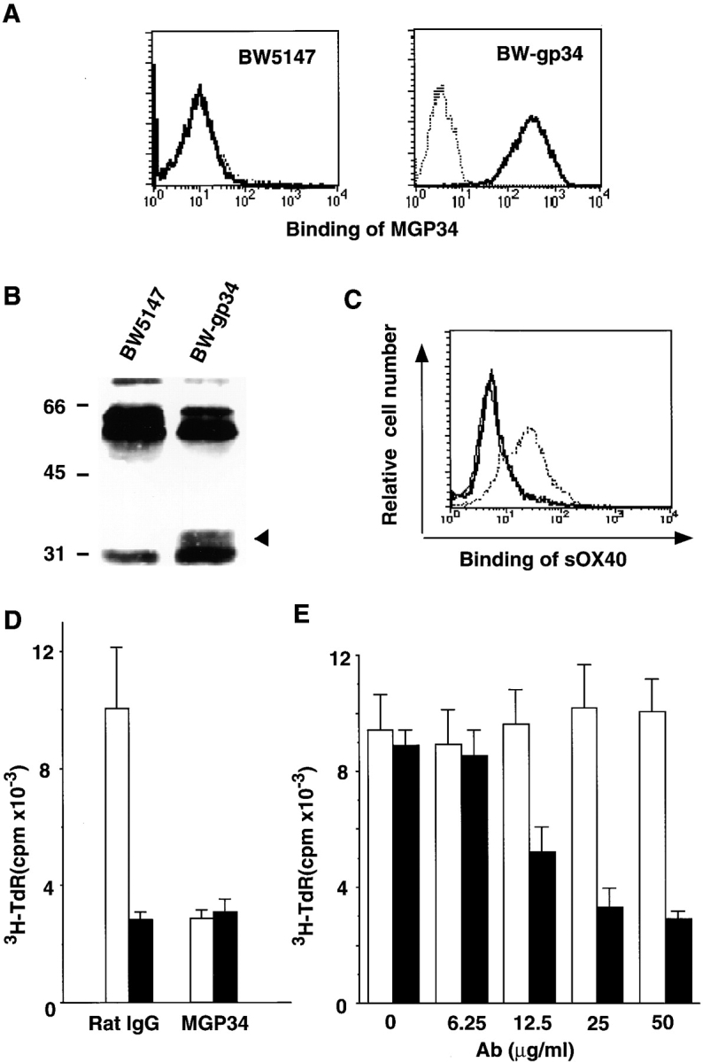
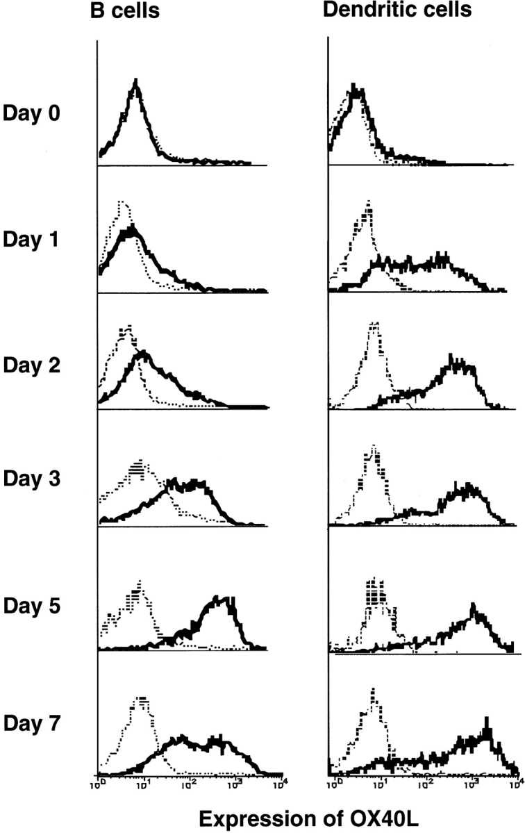
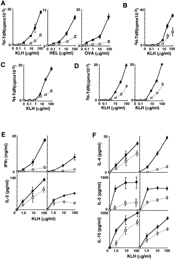
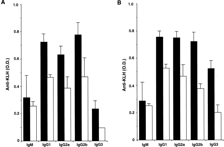
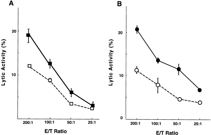
OX40 expressed on activated T cells is known to be an important costimulatory molecule on T cell activation in vitro. However, the in vivo functional significance of the interaction between OX40 and its ligand, OX40L, is still unclear. To investigate the role of OX40L during in vivo immune responses, we generated OX40L-deficient mice and a blocking anti-OX40L monoclonal antibody, MGP34. OX40L expression was demonstrated on splenic B cells after CD40 and anti-immunoglobulin (Ig)M stimulation, while only CD40 ligation was capable of inducing OX40L on dendritic cells. OX40L-deficient and MGP34-treated mice engendered apparent suppression of the recall reaction of T cells primed with both protein antigens and alloantigens and a significant reduction in keyhole limpet hemocyanin-specific IgG production. The impaired T cell priming was also accompanied by a concomitant reduction of both T helper type 1 (Th1) and Th2 cytokines. Furthermore, antigen-presenting cells (APCs) derived from the mutant mice revealed an impaired intrinsic APC function, demonstrating the importance of OX40L in both the priming and effector phases of T cell activation. Collectively, these results provide convincing evidence that OX40L, expressed on APCs, plays a critical role in antigen-specific T cell responses in vivo.


Figures

Figure 1
Figure 1
Creation of OX40L-deficient mice. (A) Structure of the wild-type and mutant OX40L alleles. The targeting construct was designed to replace the first exon (black box) with a PGKneo gene cassette. The location of the probe for hybridization, the 0.8-kb PvuII-StuI fragment, is shown (hatched box). (B) Southern blot analysis of offspring of the germline chimera. Tail DNA harvested from wild-type, OX40L+/−, and OX40L−/− mice was digested with HindIII (the location of which is depicted as H in A), electrophoresed, and probed with the radiolabeled probe. The mutant and wild-type alleles gave 10.0- and 3.0-kb hybridizing bands, respectively. (C) FACS® analysis of surface expression of OX40L. Splenic B cells from wild-type and OX40L−/− mice were stimulated with anti-CD40 plus anti-IgM, and then stained with anti–mouse OX40L mAb, MGP34.
Figure 2
Figure 2
Binding specificity and blocking potential of MGP34. (A) Specificity of MGP34 for OX40L. BW5147 parental cells (left) and BW5147 cells transfected with mouse OX40L, BW-gp34 (right), were stained with MGP34 and analyzed by flow cytometry. Dotted lines represent control stainings conducted in the presence of excess unlabeled MGP34; solid lines represent MGP34-specific staining. (B) Immunoprecipitation of mouse OX40L with MGP34. BW5147 and BW-gp34 cells were surface-labeled with biotin, lysed, and immunoprecipitated with MGP34. Arrowhead, a 34-kD precipitate. Numbers on the left indicate standard molecular sizes. (C) Competitive binding of sOX40-Fc and MGP34 to OX40L. BW-gp34 cells were preincubated with MGP34 or control IgG, and incubated with sOX40-Fc at 4°C for 30 min. The binding of the sOX40-Fc (provided by Dr. P. Baum) was visualized by anti–human Fc-FITC. Solid thick or dotted line represents sOX40-Fc binding in the presence of MGP34 or the control Ab, respectively. Solid thin line represents background staining with anti–human Fc-FITC alone. (D) Blocking effect of MGP34 on the OX40-dependent proliferation of T cells. Purified splenic T cells (105/well) were assayed for their proliferation induced by immobilized anti-CD3 in the presence of irradiated BW5147 (black bars) or BW-gp34 cells (5 × 10⁴; white bars). The cells were cultured for 72 h in the presence of 10 μg/ml of MGP34 or the control Ab, and assayed for thymidine incorporation. (E) The inhibitory effect of MGP34 is dose dependent. Purified splenic T cells (105/well) were cocultured with irradiated BW-gp34 cells (5 × 10⁴/well) in the presence of anti-CD3 at the indicated concentrations of MGP34 (black bars) or control IgG (white bars). The proliferative response was assessed at 72 h as described above.
Figure 3
Figure 3
Kinetics of OX40L expression on APCs. Splenic B cells were cultured in the presence of anti-CD40 plus anti-IgM for the indicated time periods. The cells were stained with anti-IgM–FITC, B220-PE, and biotinylated MGP34 followed by allophycocyanin-labeled streptavidin. B220+ B cells during the culture were examined for expression of OX40L. Enriched splenic dendritic cells were cultured in the presence of anti-CD40 plus anti–hamster Ab for the indicated time. The cells were stained with anti-CD11c–FITC, 33D1-PE, and biotinylated MGP34 followed by allophycocyanin-labeled streptavidin. CD11c+33D1+ dendritic cells during the culture were examined for expression of OX40L. Dotted lines represent control stainings conducted in the presence of excess unlabeled MGP34; solid lines represent MGP34-specific staining.
Figure 4
Figure 4
Impaired T cell priming and cytokine production in OX40L-deficient mice and MGP34-treated mice. (A) OX40L-deficient mice have impaired recall proliferative responses to protein antigens. OX40L-deficient (□) or wild-type (▪) mice (four per group) were immunized with KLH, HEL, or OVA in the hind footpads. 9 d after immunization, draining lymph nodes were extracted and subjected to an in vitro challenge of the various protein antigens. After culturing for 3 d, their [3H]thymidine uptake was measured. (B) Impairment of recall proliferative response of the CD4+ T cells of OX40L-deficient mice. Purified CD4+ T cells from the draining lymph nodes of the OX40L-deficient (□) or wild-type mice (▪) were assayed for their recall proliferation in response to KLH in the presence of APCs from wild-type mice. (C) Defective APC function in OX40L-deficient mice. Purified CD4+ T cells from the draining lymph nodes from wild-type mice primed with KLH were assayed for their recall proliferation in response to KLH in the presence of OX40L-deficient (□) or wild-type (▪) irradiated spleen cells used as APCs. (D) MGP34-treated mice have similarly impaired lymph node recall proliferation to KLH. C57BL/6 mice (four per group) were immunized in the hind footpads with KLH in CFA. On days 0, 3, and the 6, mice received 500 μg anti-OX40L (○) or rat IgG (•) intraperitoneally. The recall proliferative responses of the draining lymph nodes (left) were tested in the same manner as described above. Purified CD4+ T cells from the draining lymph nodes of the MGP34-treated (○) or control IgG–treated (•) mice were assayed for their recall proliferation in response to KLH in the presence of APCs from C57BL/6 mice (right). (E and F) Absence of OX40L or MGP34 treatment inhibits recall cytokine production by lymph nodes in response to KLH. Production of Th1 (E) and Th2 (F) cytokines by the draining lymph nodes of the mice in the four groups (□, ▪, ○, •) immunized with KLH and further subjected to an in vitro challenge with KLH were measured.
Figure 5
Figure 5
Impaired Ab production to KLH in OX40L-deficient mice. (A) Primary Ab responses to KLH in wild-type (black bars) and OX40L-deficient (white bars) mice were evaluated. After KLH immunization, serum was collected on day 7 for IgM and day 14 for the IgGs to examine the concentration of anti–KLH-specific Abs. (B) Secondary Ab responses to KLH in wild-type (black bars) and OX40L-deficient (white bars) mice were evaluated. Serum was collected on day 5 after the second immunization.
Figure 6
Figure 6
Suppression of CTL induction in OX40L-deficient and MGP34-treated mice. (A) OX40L-deficient (□) or wild-type (▪) mice (H-2b) were immunized intraperitoneally with splenocytes of BALB/c (H-2d) mice. On day 5, CTL activities of their splenocytes were tested with BALB/c target cells. Data shown are representative of four experiments. (B) C57BL/6 (H-2b) mice were immunized intraperitoneally with splenocytes of BALB/c (H-2d) mice. The mice were treated with 500 μg MGP34 (○) or rat IgG (•) on days 0, 2, and 4 after immunization. On day 5, CTL activities of their splenocytes were tested with BALB/c target cells. Data shown are representative of four experiments.

References

    1. Paterson D.J., Jefferies W.A., Green J.R., Brandon M.R., Corthesy P., Puklavec M., Williams A.F. Antigens of activated rat T lymphocytes including a molecule of 50,000 M r detected only on CD4 positive T blasts. Mol. Immunol. 1987;24:1281–1290. - PubMed
    1. Mallett S., Fossum S., Barclay A.N. Characterization of the MRC OX40 antigen of activated CD4 positive T lymphocytes—a molecule related to nerve growth factor receptor. EMBO (Eur. Mol. Biol. Organ.) J. 1990;9:1063–1068. - PMC - PubMed
    1. Al-Shamkhani A., Birkeland M.L., Puklavec M., Brown M.H., James W., Barclay A.N. OX40 is differentially expressed on activated rat and mouse T cells and is the sole receptor for the OX40 ligand. Eur. J. Immunol. 1996;26:1695–1699. - PubMed
    1. Calderhead D.M., Buhlmann J.E., Van der Eetwegh A.J.M., Claasen E., Noelle R.J., Fell H.P. Cloning of mouse Ox40a T cell activation marker that may mediate T-B interactions. J. Immunol. 1993;151:5261–5271. - PubMed
    1. Baum P.R., Gayle R.B., Ramsdell F., Srinivasan S., Sorensen R.A., Watson M.L., Seldin M.F., Baker E., Sutherland G.R., Clifford K.N. Molecular characterization of murine and human OX-40/OX-40 ligand systemsidentification of a human OX-40 ligand as the HTLV-1-regulated protein gp34. EMBO (Eur. Mol. Biol. Organ.) J. 1994;13:3992–4001. - PMC - PubMed

Publication types

MeSH terms