. 2000 Jan 17;191(2):375-80.
doi: 10.1084/jem.191.2.375.

Critical contribution of OX40 ligand to T helper cell type 2 differentiation in experimental leishmaniasis

Affiliations

Critical contribution of OX40 ligand to T helper cell type 2 differentiation in experimental leishmaniasis

H Akiba et al. J Exp Med. .

Abstract

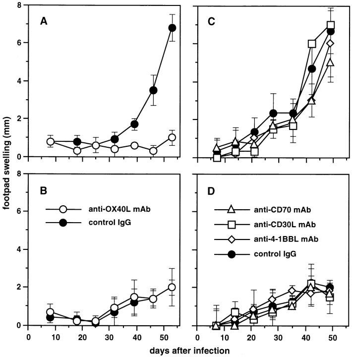
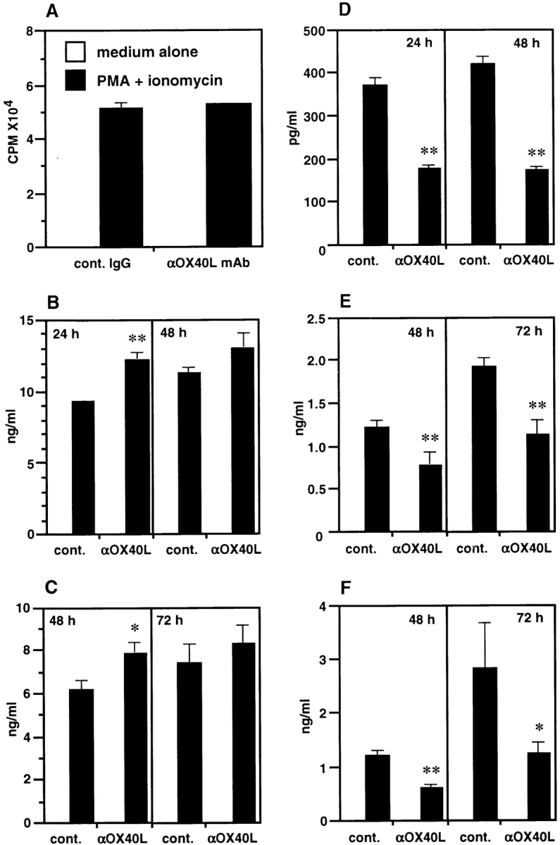
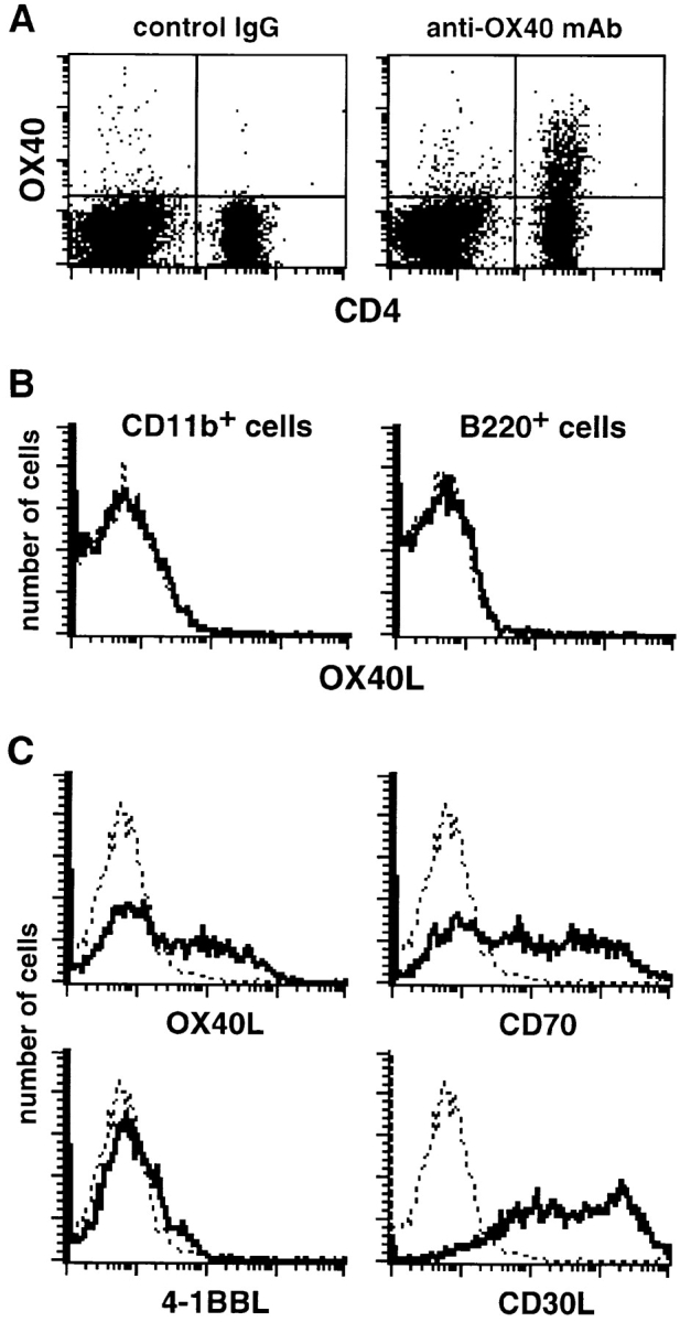
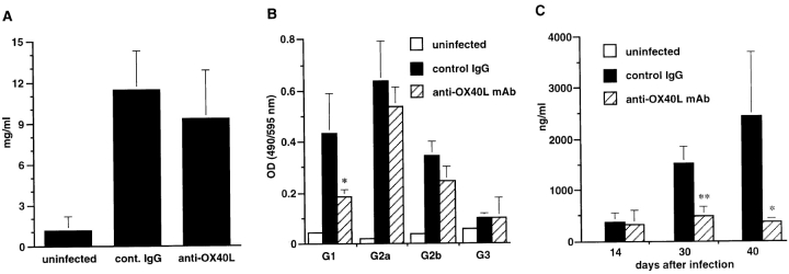
Infection of inbred mouse strains with Leishmania major is a well characterized model for analysis of T helper (Th)1 and Th2 cell development in vivo. In this study, to address the role of costimulatory molecules CD27, CD30, 4-1BB, and OX40, which belong to the tumor necrosis factor receptor superfamily, in the development of Th1 and Th2 cells in vivo, we administered monoclonal antibody (mAb) against their ligands, CD70, CD30 ligand (L), 4-1BBL, and OX40L, to mice infected with L. major. Whereas anti-CD70, anti-CD30L, and anti-4-1BBL mAb exhibited no effect in either susceptible BALB/c or resistant C57BL/6 mice, the administration of anti-OX40L mAb abrogated progressive disease in BALB/c mice. Flow cytometric analysis indicated that OX40 was expressed on CD4(+) T cells and OX40L was expressed on CD11c(+) dendritic cells in the popliteal lymph nodes of L. major-infected BALB/c mice. In vitro stimulation of these CD4(+) T cells showed that anti-OX40L mAb treatment resulted in substantially reduced production of Th2 cytokines. Moreover, this change in cytokine levels was associated with reduced levels of anti-L. major immunoglobulin (Ig)G1 and serum IgE. These results indicate that anti-OX40L mAb abrogated progressive leishmaniasis in BALB/c mice by suppressing the development of Th2 responses, substantiating a critical role of OX40-OX40L interaction in Th2 development in vivo.


Figures

Figure 1
Figure 1
Effect of anti-OX40L, -CD70, -CD30L, and –4-1BBL mAb on the course of L. major infection. BALB/c mice (A and C) or C57BL/6 mice (B and D) were infected with 5 × 10⁶ stationary phase promastigotes subcutaneously in the hind footpad. Mice were treated with 300 μg of anti-OX40L mAb (○, A and B), anti-CD70 (▵), anti-CD30L (□), or anti–4-1BBL (⋄) mAb (C and D) or control IgG (•, A–D) intraperitoneally at the time of infection and subsequently twice per week until the end of experiments. Net footpad swelling was measured by subtracting the thickness of the uninfected footpad from that of the infected footpad. Results are expressed as mean ± SD of six mice in each group. Similar results were obtained in two independent experiments.
Figure 3
Figure 3
Functional phenotype of CD4+ T cells in anti-OX40L–treated BALB/c mice. CD4+ T cells were purified from popliteal LNs of L. major–infected BALB/c mice that were treated with anti-OX40L mAb or control IgG 50 d after infection and stimulated with PMA and ionomycin for 24–72 h. (A) Proliferative response at 48 h was assessed by pulsing the cultures with 0.5 μCi/well of [3H]thymidine for the last 7 h. Production of IL-2 (B), IFN-γ (C), IL-4 (D), IL-10 (E), and IL-13 (F) in culture supernatants at the indicated periods was measured by ELISA. Results are expressed as mean ± SD of triplicate cultures. Significant differences between control IgG and anti-OX40L mAb treatment are indicated by asterisks (*P < 0.05; **P < 0.01). Similar results were obtained in three independent experiments.
Figure 2
Figure 2
Expression of OX40 and OX40L on popliteal LN cells from L. major–infected BALB/c mice at day 40 after infection. (A) OX40 is expressed on CD4+ T cells. Popliteal LN cells proximal to the footpad lesions of L. major–infected BALB/c mice were double stained with FITC-labeled anti-CD4 mAb and biotinylated anti-OX40 mAb or control IgG followed by PE-labeled streptavidin. (B) OX40L is not expressed on CD11b+ Mφ and B220+ B cells. Popliteal LN cells were double stained with FITC-labeled anti-CD11b or anti-B220 mAb and biotinylated anti-OX40L mAb or control IgG followed by PE-labeled streptavidin. The histograms show staining of electronically gated CD11b+ or B220+ cells. Bold line shows staining with anti-OX40L mAb, and dotted line shows background staining with control IgG. (C) Expression of OX40L, CD70, 4-1BBL, and CD30L on CD11c+ DCs. Low density cells from popliteal LNs were double stained with FITC-labeled anti-CD11c mAb and biotinylated anti-OX40L, CD70, 4-1BBL, or CD30L mAb or control IgG followed by PE-labeled streptavidin. The histograms show staining of electronically gated CD11c+ cells. Bold line shows staining with mAb against the indicated molecules, and dotted line shows background staining with control IgG.
Figure 4
Figure 4
Effect of anti-OX40L mAb treatment on serum IgG Ab and IgE in L. major–infected BALB/c mice. (A) Serum total IgG 40 d after infection was measured by sandwich ELISA. Results are expressed as mean ± SD of six mice in each group. (B) L. major–specific IgG Ab isotypes were determined with serum 40 d after infection against SLAs by isotype-specific ELISA. Data represent OD obtained at a 500-fold dilution of serum and are expressed as mean ± SD of six mice in each group. (C) Serum IgE at 14, 30, and 40 d after infection was measured by IgE-specific sandwich ELISA. Results are expressed as mean ± SD of six mice in each group. Significant differences between control IgG and anti-OX40L mAb treatment are indicated by asterisks (*P < 0.05; **P < 0.01). Similar results were obtained in two independent experiments.

References

    1. O'Garra A. Cytokines induce the development of functionally heterogeneous T helper cell subsets. Immunity. 1998;8:275–283. - PubMed
    1. Kuchroo V.K., Das M.P., Brown J.A., Ranger A.M., Zamvil S.S., Sobel R.A., Weiner H.L., Nabavi N., Glimcher L.H. B7-1 and B7-2 costimulatory molecules activate differentially the Th1/Th2 developmental pathwaysapplication to autoimmune disease therapy. Cell. 1995;80:707–718. - PubMed
    1. Smith C.A., Farrah T., Goodwin R.G. The TNF receptor superfamily of cellular and viral proteinsactivation, costimulation, and death. Cell. 1994;76:959–962. - PubMed
    1. Gruss H.J., Dower S.K. Tumor necrosis factor ligand superfamilyinvolvement in the pathology of malignant lymphomas. Blood. 1995;85:3378–3404. - PubMed
    1. Sugita K., Torimoto Y., Nojima Y., Daley J.F., Schlossman S.F., Morimoto C. The 1A4 molecule (CD27) is involved in T cell activation. J. Immunol. 1991;147:1477–1483. - PubMed

Publication types

MeSH terms

Substances