. 2000 Jan;11(1):1-11.
doi: 10.1091/mbc.11.1.1.

Regulation of mitotic inhibitor Mik1 helps to enforce the DNA damage checkpoint

Affiliations

Regulation of mitotic inhibitor Mik1 helps to enforce the DNA damage checkpoint

B A Baber-Furnari et al. Mol Biol Cell. 2000 Jan.

Abstract

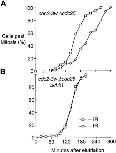
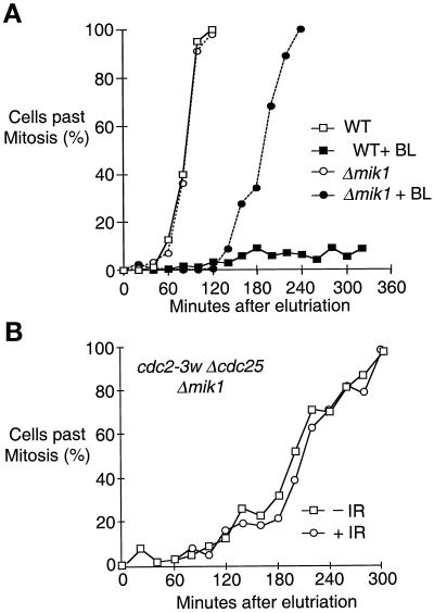
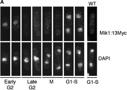
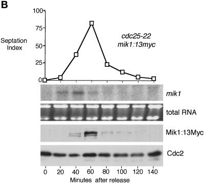
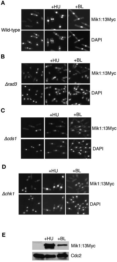
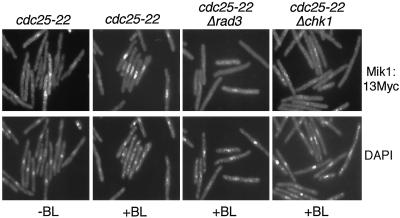
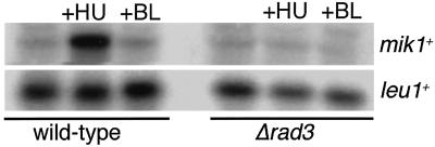
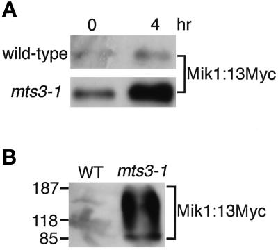
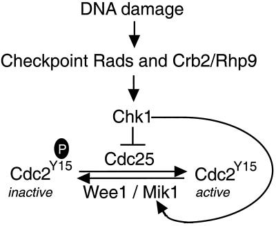
The protein kinase Chk1 enforces the DNA damage checkpoint. This checkpoint delays mitosis until damaged DNA is repaired. Chk1 regulates the activity and localization of Cdc25, the tyrosine phosphatase that activates the cdk Cdc2. Here we report that Mik1, a tyrosine kinase that inhibits Cdc2, is positively regulated by the DNA damage checkpoint. Mik1 is required for checkpoint response in strains that lack Cdc25. Long-term DNA damage checkpoint arrest fails in Deltamik1 cells. DNA damage increases Mik1 abundance in a Chk1-dependent manner. Ubiquitinated Mik1 accumulates in a proteasome mutant, which indicates that Mik1 normally has a short half-life. Thus, the DNA damage checkpoint might regulate Mik1 degradation. Mik1 protein and mRNA oscillate during the unperturbed cell cycle, with peak amounts detected around S phase. These data indicate that regulation of Mik1 abundance helps to couple mitotic onset to the completion of DNA replication and repair. Coordinated negative regulation of Cdc25 and positive regulation of Mik1 ensure the effective operation of the DNA damage checkpoint.


Figures

Figure 1
Figure 1
Chk1-dependent mitotic delay caused by ionizing irradiation in cdc2-3w Δcdc25 cells. Synchronous populations of cdc2-3w Δcdc25 (GL192) or cdc2-3w Δcdc25 Δchk1 (NR1976) cells were obtained by centrifugal elutriation. Cultures were exposed to 200 Gy of ionizing radiation (+IR) or mock treated (−IR). Progression through mitosis was determined by microscopic observation.
Figure 2
Figure 2
GST–Chk1 overproduction arrests division in the absence of Wee1 and Cdc25. A wee1-50 Δcdc25 nmt1:GST-chk1+ (BF2404) strain was incubated on medium that repressed (off) or induced (on) GST–Chk1 production. Cells were photographed after 48 h at 35°C.
Figure 3
Figure 3
Deletion of mik1+ impairs the DNA damage checkpoint. (A) Synchronous cultures of early G2 wild-type (PR109) or Δmik1 (PR712) cells were produced by centrifugal elutriation and exposed to the radiomimetic drug bleomycin (BL) at a concentration of 2.5 mU/ml or mock treated. (B) Synchronous cultures of early G2 cdc2-3w Δcdc25 Δmik1 (NR1977) cells were exposed to 200 Gy of ionizing irradiation (+IR) or mock treated (−IR). Note that NR1977 grows slowly relative to cdc2-3w Δcdc25 (GL192) shown in Figure 1A; thus, these experiments cannot be compared directly.
Figure 4
Figure 4
Nuclear staining of Mik1 during S phase and early G2 corresponds to increased Mik1 mRNA and protein. (A) A synchronous population of mik1+:13Myc (AL2405) cells in early G2 phase was produced by centrifugal elutriation and processed to detect Mik1:13Myc or nuclei with the DNA stain DAPI. The approximate phase of the cell cycle is indicated. Nuclear Mik1 was detected during S phase and early G2. Cytoplasmic staining was not significantly different from that of the untagged wild-type (WT) strain. (B) Synchronous cdc25-22 mik1+:13Myc (BF2409) cells in late G2 were obtained by incubation at restrictive temperature (36°C) for 4 h. Division arrest was relieved by decreasing the temperature to 25°C. Northern blot analysis revealed that mik1 mRNA peaked shortly after the division release (40 min), whereas immunoblot studies showed that Mik1 protein abundance peaked soon thereafter (60 min). These time points correspond approximately to S or early G2 phase. Loading controls were total RNA for the Northern blot and Cdc2 protein for the immunoblot.
Figure 4
Figure 4
Nuclear staining of Mik1 during S phase and early G2 corresponds to increased Mik1 mRNA and protein. (A) A synchronous population of mik1+:13Myc (AL2405) cells in early G2 phase was produced by centrifugal elutriation and processed to detect Mik1:13Myc or nuclei with the DNA stain DAPI. The approximate phase of the cell cycle is indicated. Nuclear Mik1 was detected during S phase and early G2. Cytoplasmic staining was not significantly different from that of the untagged wild-type (WT) strain. (B) Synchronous cdc25-22 mik1+:13Myc (BF2409) cells in late G2 were obtained by incubation at restrictive temperature (36°C) for 4 h. Division arrest was relieved by decreasing the temperature to 25°C. Northern blot analysis revealed that mik1 mRNA peaked shortly after the division release (40 min), whereas immunoblot studies showed that Mik1 protein abundance peaked soon thereafter (60 min). These time points correspond approximately to S or early G2 phase. Loading controls were total RNA for the Northern blot and Cdc2 protein for the immunoblot.
Figure 5
Figure 5
DNA replication and damage checkpoints stimulate nuclear accumulation of Mik1. Asynchronous populations of strains that contained the mik1+:13Myc allele were incubated in 10 mM hydroxyurea (+HU) or 2.5 mU/ml bleomycin (+BL) for 4 h. Cells were processed to reveal Mik1:13Myc or DNA (DAPI). (A) Wild type (AL2405); (B) Δrad3 (BF2406); (C) Δcds1 (BF2408); (D) Δchk1 (BF2407). (E) Immunoblot analysis of wild-type (AL2405) cells exposed to HU or BL as described above.
Figure 6
Figure 6
Chk1-dependent nuclear accumulation of Mik1 is not a consequence of the G2 arrest. Cultures of cdc25-22 mik1+:13Myc (BF2409), cdc25-22 rad3::ura4+ mik1:13Myc (BF2442), or cdc25-22 mik1+:13Myc Δchk1 (BF2410) cells were shifted to restrictive temperature (36°C). Thirty minutes later, cells were treated with 2.5 mU/ml bleomycin (+BL) or mock treated (−BL). After another 3.5-h incubation at 36°C, cells were processed to detect Mik1:13Myc or DNA (DAPI).
Figure 7
Figure 7
Regulation of mik1 mRNA in checkpoint-arrested cells. Wild-type (AL2405) or Δrad3 (BF2406) cells were incubated in 10 mM hydroxyurea (+HU) or 2.5 mU/ml bleomycin (+BL) for 4 h. RNA was harvested and probed in a Northern blot with mik1 or leu1 probes. PhosphorImager analysis indicated that mik1 mRNA increased ∼3.5-fold in the HU-treated wild-type cells relative to all the other samples.
Figure 8
Figure 8
Accumulation of ubiquitinated Mik1 in proteasome mutant. (A) Wild-type (AL2405) or mts3-1 (NB2411) cells that expressed Mik1:13Myc from the mik1+ locus were incubated for 4 h at 35.5°C. Anti-myc immunoblot analysis was performed with cell extracts. (B) The same strains were transformed with pREP1-His6-ubiquitin and induced to express His6-ubiquitin before incubation at 4 h at 35.5°C. Samples were processed to purify proteins that were covalently linked to His6-ubiquitin followed by immunoblot analysis with anti-Myc antibody.
Figure 9
Figure 9
Model of the DNA damage checkpoint in fission yeast.

References

    1. al-Khodairy F, Carr AM. DNA repair mutants defining G2 checkpoint pathways in Schizosaccharomyces pombe. EMBO J. 1992;11:1343–1350. - PMC - PubMed
    1. al-Khodairy F, Fotou E, Sheldrick KS, Griffiths DJ, Lehmann AR, Carr AM. Identification and characterization of new elements involved in checkpoint and feedback controls in fission yeast. Mol Biol Cell. 1994;5:147–160. - PMC - PubMed
    1. Blasina A, Van de Weyer I, Laus MC, Luyten WHML, Parker AE, McGowan CH. A human homolog of the checkpoint kinase Cds1 directly inhibits Cdc25. Curr Biol. 1999;9:1–10. - PubMed
    1. Boddy MN, Furnari B, Mondesert O, Russell P. Replication checkpoint enforced by kinases Cds1 and Chk1. Science. 1998;280:909–912. - PubMed
    1. Booher R, Beach D. Involvement of cdc13+ in mitotic control in Schizosaccharomyces pombe: possible interaction of the gene product with microtubules. EMBO J. 1988;7:2321–2327. - PMC - PubMed

Publication types

MeSH terms