. 2000 Jan;11(1):171-82.
doi: 10.1091/mbc.11.1.171.

Organization of the yeast Golgi complex into at least four functionally distinct compartments

Affiliations
Free PMC article

Organization of the yeast Golgi complex into at least four functionally distinct compartments

W T Brigance et al. Mol Biol Cell. 2000 Jan.
Free PMC article

Abstract

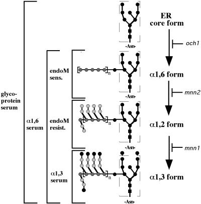
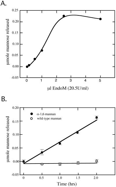
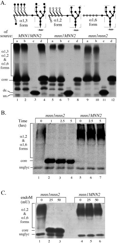
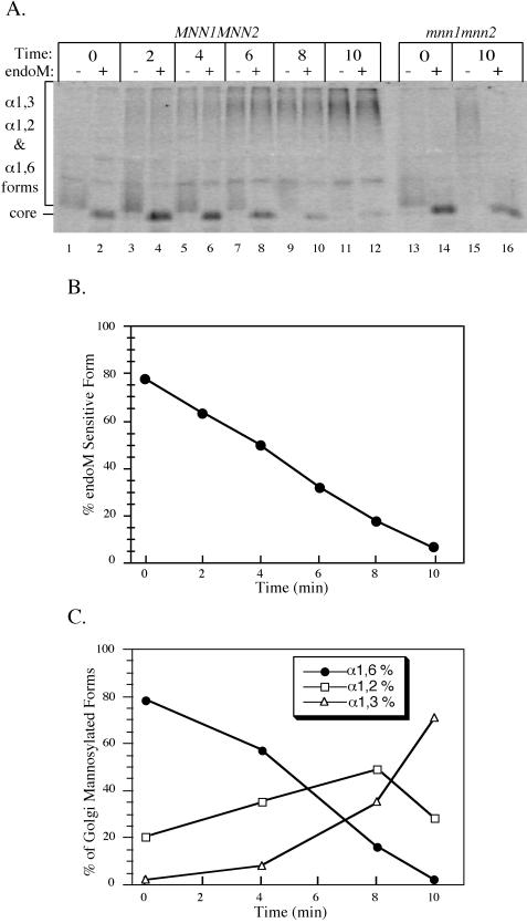
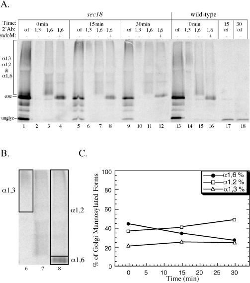
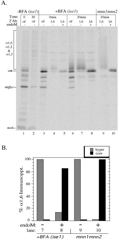
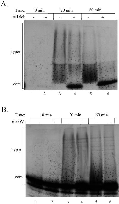
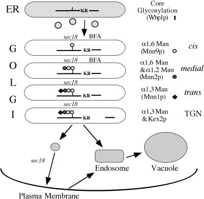
Pro-alpha-factor (pro-alphaf) is posttranslationally modified in the yeast Golgi complex by the addition of alpha1,6-, alpha1,2-, and alpha1,3-linked mannose to N-linked oligosaccharides and by a Kex2p-initiated proteolytic processing event. Previous work has indicated that the alpha1,6- and alpha1,3-mannosylation and Kex2p-dependent processing of pro-alphaf are initiated in three distinct compartments of the Golgi complex. Here, we present evidence that alpha1,2-mannosylation of pro-alphaf is also initiated in a distinct Golgi compartment. Linkage-specific antisera and an endo-alpha1,6-D-mannanase (endoM) were used to quantitate the amount of each pro-alphaf intermediate during transport through the Golgi complex. We found that alpha1,6-, alpha1,2-, and alpha1,3-mannose were sequentially added to pro-alphaf in a temporally ordered manner, and that the intercompartmental transport factor Sec18p/N-ethylmaleimide-sensitive factor was required for each step. The Sec18p dependence implies that a transport event was required between each modification event. In addition, most of the Golgi-modified pro-alphaf that accumulated in brefeldin A-treated cells received only alpha1,6-mannosylation as did approximately 50% of pro-alphaf transported to the Golgi in vitro. This further supports the presence of an early Golgi compartment that houses an alpha1,6-mannosyltransferase but lacks alpha1,2-mannosyltransferase activity in vivo. We propose that the alpha1,6-, alpha1,2-, and alpha1,3-mannosylation and Kex2p-dependent processing events mark the cis, medial, trans, and trans-Golgi network of the yeast Golgi complex, respectively.


Figures

Figure 1
Figure 1
Method for defining the biosynthetic intermediates of pro-αf. Our nomenclature for the pro-αf biosynthetic intermediates is shown to the right, and the method for defining the intermediates is to the left of the structures. Briefly, all forms are recognized by the primary antiserum. The α1,6-linkage-specific serum recognizes all Golgi mannosylated forms, whereas the α1,3-linkage-specific serum only recognizes the terminal α1,3-mannosylated form. EndoM-sensitive (sens.) pro-αf forms carry unbranched α1,6-mannan chains, whereas endoM-resistant (resist.) forms have been branched with α1,2-mannose. Each step is defined genetically by mutants lacking each mannose modification as indicated.
Figure 2
Figure 2
Activity and specificity of purified endoM against a crude mannan substrate. Release of reduced mannose from a mannan substrate was measured using the Nelson–Somogyi colorimetric assay. (A) 250 μg of mannan substrate (per reaction) prepared from strain TH2-10D (mnn1 mnn2) were incubated with an increasing amount of purified endoM for 2 h at 50°C. The reaction was saturated with ∼50 mU of enzyme. (B) 250 μg of mannan prepared from mnn1 mnn2 cells (closed circles) or wild-type cells (open circles) were incubated with 40 mU of endoM for the time points indicated. The mnn1 mnn2 strain produces α1,6-mannan lacking α1,2- or α1,3-linked mannose.
Figure 3
Figure 3
Activity and specificity of endoM preparations against immunoprecipitated pro-αf. (A) MNN1 MNN2 (TBY130), mnn1 MNN2 (TBY131), and mnn1 mnn2 (TBY132) strains were labeled with 35S-amino acids for 10 min at 20°C and no chase. N-linked oligosaccharides produced from these yeast strains are represented above each genotype. Pro-αf was recovered from the cell lysates by immunoprecipition and was split into four equal samples. One sample was left untreated (lanes marked a), and the other samples were treated with the following preparations: (b) endoM buffer (mock), (c) ammonium sulfate-purified endoM (50mU), and (d) DEAE-purified endoM (50 mU). All subsequent enzyme incubations are with DEAE-purified endoM. (B and C) Pro-αf was immunoprecipitated from mnn1 and mnn1 mnn2 yeast strains labeled at 20°C for 10 min and chased for 7 min. (B) Samples were split and treated with 50 mU of endoM for the indicated times. (C)Samples were split and treated with the indicated amount of endoM. de., deglycosylated pro-αf; un., unglycosylated pro-αf. Note that the contaminating endoglycosidase apparent in lanes marked C produces a deglycosylated pro-αf with a slightly slower mobility than the unglycosylated form. This suggests cleavage between the N-acetylglucosamines leaving behind one sugar per N-linked oligosaccharide. Core indicates the ER pro-αf form carrying the oligosaccharide shown within the dashed box. All three experiments were electorphoresed on 15% SDS-polyacrylamide gels with B and C run for shorter times than A.
Figure 4
Figure 4
Sequential modification of pro-αf in the Golgi complex. (A) kex2Δ cells (TBY130) were labeled at 15°C for 5 min and chased for the times indicated. Pro-αf was immunoprecipitated first with αf serum and then with α1,6-linkage serum and split in half for treatment with or without endoM. mnn1 mnn2kex2Δ (TBY131) cells were also labeled and pro-αf analyzed as described above. (B) Quantitation of the data in A was carried out using a phosphorimager. Forms resistant to endoM are a mixture of α1,2- and α1,3-mannosylated pro-αf, whereas sensitive forms are only α1,6-mannosylated. (C) The kex2Δ strain TBY130 was labeled and chased as above. The intermediate forms were quantified as described in MATERIALS AND METHODS section and shown in Figures 1 and 5. The data shown are from a single experiment but are representative of data obtained in three independent experiments.
Figure 5
Figure 5
Sec18p is required for the formation and consumption of each pro-αf intermediate form. Wild-type (SEY6210) and sec18 (TBY102) cells were labeled at 20°C for 7 min and shifted to 37°C for a 30-min chase. The secondary immunoprecipitations and endoM treatment were performed as described in MATERIALS AND METHODS section. All samples (1.5 OD equivalents) were electrophoresed in 15% polyacrylamide gels and analyzed by phosporimaging. (B) The area of the lanes used to quantitate the α1,6-, α1,2-, and α1,3-mannosylated forms from the 15-min time point is boxed. (C) Quantitation of α1,6-, α1,2-, and α1,3-mannosylated pro-af forms at each time point for the sec18 samples. Each value is the average from three independent experiments.
Figure 6
Figure 6
BFA-treated cells accumulate core glycosylated and α1,6-mannosylated pro-αf. BFA-sensitive cells (ise1) were pretreated for 10 min at 20°C with or without 75 μg/ml BFA, labeled for 7 min, and chased for 30 min. Pro-αf was recovered from each sample by immunoprecipitation and subjected to a second immunoprecipitation with αf- or α1,6-linkage serum. The α1,6 immunoprecipitates were incubated with or without endoM before electrophoresis in a 15% polyacrylamide gel. Pro-αf was also recovered from labeled mnn1 mnn2 cells for a positive control for the endoM treatment. unglyc, unglycosylated pro-αf; mαf, mature αf. (B) Quantitation of the data shown in lanes 7–10 of A. In this experiment, a small amount of the core glycosylated pro-αf contaminated the α1,6 immunoprecipitations (lanes 4 and 7) and was subtracted as background from the BFA-treated samples shown in B.
Figure 7
Figure 7
ER to Golgi transport assay results in both α1,6-mannosylated and α1,2-mannosylated forms of pro-αf. Semi-intact yeast cells were incubated with cytosol, 35S-labeled prepro-αf, and ATP for the times indicated. Samples from each time point were removed and immunoprecipitated with either α1,6-linkage serum (A) or concanavalin A-Sepharose (B), split evenly, and treated with or without endoM.
Figure 8
Figure 8
Compartmental organization of the yeast Golgi complex. (see DISCUSSION for explanation).

References

    1. Baker D, Hicke L, Rexach M, Schleyer M, Schekman R. Reconstitution of SEC gene product-dependent intercompartmental protein transport. Cell. 1988;54:335–344. - PubMed
    1. Barlowe C, Orci L, Yeung T, Hosobuchi M, Hamamoto S, Salama N, Rexach MF, Ravazzola M, Amherdt M, Schekman R. COPII: a membrane coat formed by Sec proteins that drive vesicle budding from the endoplasmic reticulum. Cell. 1994;77:895–907. - PubMed
    1. Bevan A, Brenner C, Fuller RS. Quantitative assessment of enzyme specificity in vivo: P2 recognition by Kex2 protease defined in a genetic system. Proc Natl Acad Sci USA. 1998;95:10384–10389. - PMC - PubMed
    1. Bussey H. Proteases and the processing of precursors to secreted proteins in yeast. Yeast. 1988;4:17–26. - PubMed
    1. Dubois M, Gilles KA, Hamilton JK, Rebers PA, Smith F. Colorimetic method for determination of sugars and related substances. Anal Chem. 1956;28:350–356.

Publication types

MeSH terms