. 2000 Jan;11(1):227-39.
doi: 10.1091/mbc.11.1.227.

ATPase-defective mammalian VPS4 localizes to aberrant endosomes and impairs cholesterol trafficking

Affiliations
Free PMC article

ATPase-defective mammalian VPS4 localizes to aberrant endosomes and impairs cholesterol trafficking

N Bishop et al. Mol Biol Cell. 2000 Jan.
Free PMC article

Abstract

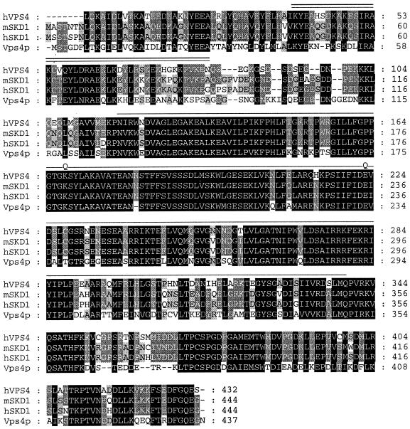
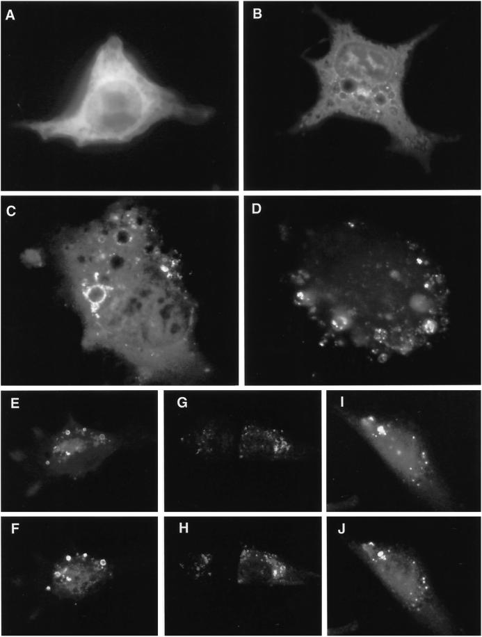
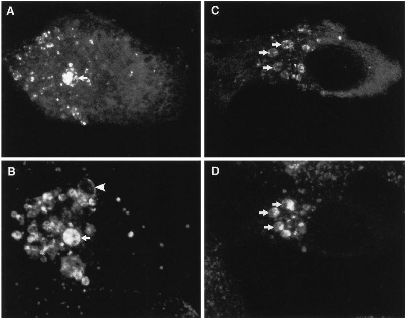
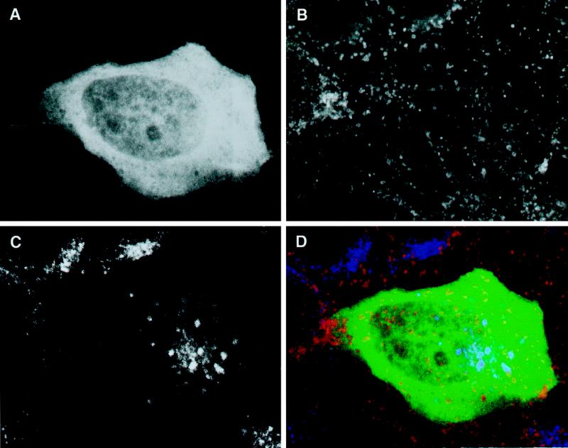
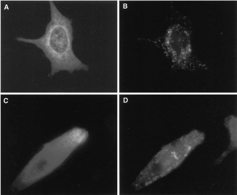
The yeast vacuolar sorting protein Vps4p is an ATPase required for endosomal trafficking that couples membrane association to its ATPase cycle. To investigate the function of mammalian VPS4 in endosomal trafficking, we have transiently expressed wild-type or ATPase-defective human VPS4 (hVPS4) in cultured cells. Wild-type hVPS4 was cytosolic, whereas a substantial fraction of hVPS4 that was unable to either bind or hydrolyze ATP was localized to membranes, including those of specifically induced vacuoles. Vacuoles were exclusively endocytic in origin, and subsets of enlarged vacuoles stained with markers for each stage of the endocytic pathway. Sorting of receptors from the early endosome to the recycling compartment or to the trans-Golgi network was not significantly affected, and no mutant hVPS4 associated with these compartments. However, many hVPS4-induced vacuoles were substantially enriched in cholesterol relative to the endosomal compartments of untransfected cells, indicating that expression of mutant hVPS4 gives rise to a kinetic block in postendosomal cholesterol sorting. The phenotype described here is largely consistent with the defects in vacuolar sorting associated with class E vps mutants in yeast, and a role for mammalian VPS4 is discussed in this context.


Figures

Figure 1
Figure 1
Characterization of human VPS4. The amino acid sequence of human VPS4 was compared with those of human and mouse SKD1 and yeast Vps4p. Identical amino acids are outlined in black. Those amino acids whose identity is conserved only between three proteins are outlined in dark gray. Light gray indicates residues where identity is limited to two proteins. The location of the N-terminal coiled coil–forming region that was deleted for some experiments is indicated by a double overline, and that of the AAA domain is indicated by a single line. Point mutations used in this study are also shown. The sequence data for hVPS4 are available from GenBank under accession number AF038960.
Figure 2
Figure 2
Expression of GFP-tagged hVPS4 in cultured NRK cells. (A) Wild-type GFP-hVPS4 was transiently expressed in NRK cells and visualized by conventional fluorescence microscopy. (B) As in A, but with cells expressing GFP-hVPS4(KQ), an ATP binding-defective mutant. (C) As in A, but with cells expressing GFP-hVPS4(EQ), an ATP hydrolysis-defective mutant. (D) Cells expressing GFP-hVPS4(EQ) were treated with 0.01% (wt/vol) saponin immediately before fixation. NRK cells were cotransfected with GFP-hVPS4(EQ) and mSKD1-myc/His6(KQ) (E and F), hVPS4(EQ) and mSKD1-myc/His6(wt) (G and H), or hVPS4(KQ) and mSKD1-myc/His6(wt) (I and J). Cells were visualized by direct fluorescence microscopy (A–D, E, G, and I) or by indirect fluorescence microscopy using anti-myc antibody (F, H, and J).
Figure 3
Figure 3
Human VPS4(EQ) binds to aberrant late endosomal compartments. NRK cells transfected with GFP-hVPS4(EQ) and permeabilized before fixation were visualized by direct confocal fluorescence microscopy (A) or indirectly using antibody against lgp120 and TRITC-conjugated secondary antibody (B). Arrows indicate lgp120-positive compartments that contain GFP-hVPS4(EQ). The arrowhead indicates an lgp120-positive compartment which lacks GFP-hVPS4(EQ). Alternatively, cells transfected with GFP-hVPS4(EQ) were visualized by direct confocal fluorescence microscopy (C) or indirectly using antibody against M6PR (D). Arrows indicate examples of M6PR-positive compartments that contain GFP-hVPS4(EQ).
Figure 4
Figure 4
Distribution of lgp120 and M6PR in cells transfected with GFP-hVPS4(wt). NRK cells transfected with GFP-hVPS4(wt) were visualized directly (A; green) or using anti-lgp120 (B; red) or anti-M6PR (C; blue). The merged image (D) shows the limited codistribution of lgp120 and M6PR. Note the similar distributions of these markers in transfected and neighboring untransfected cells.
Figure 5
Figure 5
Expression of hVPS4(EQ) gives rise to aberrant early endosomes. BHK cells were transfected with GFP-hVPS4(EQ) (A–C) or GFP-hVPS4(wt) (D–F) and visualized directly (A and D; green) or after incubation with anti-TfR (B and E; red). Merged images are shown (C and F). Arrows (A and B) provide examples of where GFP-hVPS4(EQ) and TfR colocalize. Note the change in TfR distribution in cells transfected with GFP-hVPS4(EQ) compared with GFP-hVPS4(wt) or untransfected cells. Cells in A–C were permeabilized with saponin before fixation.
Figure 6
Figure 6
Human VPS4 expression does not cause mixing of lgp120 and M6PR compartments. NRK cells expressing GFP-hVPS4(EQ) were treated with saponin before fixation and examined by direct fluorescence microscopy for GFP (A; green) or by indirect fluorescence microscopy for lgp120 (B; red) and M6PR (C; blue). (D) Merged image. All images are from a single confocal slice. GFP-positive structures that label with M6PR are indicated by arrows. Examples of lgp120-positive structures that lack GFP are indicated by arrowheads.
Figure 7
Figure 7
Human VPS4 expression does not affect sorting of TGN38 or transferrin receptor. NRK cells expressing GFP-hVPS4(wt) (A and C) or GFP-hVPS4(EQ) (B and D) were treated with saponin before fixation and examined by confocal fluorescence microscopy for GFP (all samples), TfR (C and D), and TGN38 (A and B).
Figure 8
Figure 8
Expression of ATPase-defective hVPS4 affects cholesterol sorting. NRK cells were transfected with GFP-hVPS4(KQ) (A–H) or GFP-hVPS4(wt) (I–L) and examined by conventional fluorescence microscopy for GFP (green; A, E, and I), M6PR (red; B), lgp120 (red; F and J), and cholesterol (blue, filipin staining; C, G, and K). Merged images (D, H, and L) are shown. Examples of vacuoles that costain for GFP, marker, and filipin are indicated by arrows. Note that exposure conditions for filipin staining were identical in all samples. No increase in filipin staining was observed in the cell transfected with GFP-hVPS4(wt) relative to the neighboring untransfected cell.
Figure 9
Figure 9
The coiled coil domain of hVPS4 is required for endosome localization and formation of aberrant compartments. NRK cells (A and B) or BHK cells (C and D) were transfected with GFP-hVPS4(EQ/ΔCC) and examined for GFP (A and C), lgp120 (B), or TfR (D).

References

    1. Adams MD, Dubnick M, Kerlavage AR, Moreno R, Kelley JM, Utterback TR, Nagle JW, Fields C, Venter JC. Sequence identification of 2375 human brain genes. Nature. 1992;355:632–634. - PubMed
    1. Babst M, Sato TK, Banta LM, Emr SD. Endosomal transport function in yeast requires a novel AAA-type ATPase, Vps4p. EMBO J. 1997;16:1820–1831. - PMC - PubMed
    1. Babst M, Wendland B, Estapa EJ, Emr SD. The Vps4p AAA ATPase regulates membrane association of a Vps protein complex required for normal endosome function. EMBO J. 1998;17:2982–2993. - PMC - PubMed
    1. Bright NA, Reaves BJ, Mullock BM, Luzio JP. Dense core lysosomes can fuse with late endosomes and are reformed from the resultant hybrid organelles. J Cell Sci. 1997;110:2027–2040. - PubMed
    1. Bryant NJ, Stevens TH. Vacuole biogenesis in Saccharomyces cerevisiae: protein transport pathways to the yeast vacuole. Microbiol Mol Biol Rev. 1998;62:230–247. - PMC - PubMed

Publication types

MeSH terms

Substances