. 2000 Jan 18;97(2):571-6.
doi: 10.1073/pnas.97.2.571.

Acceleration of oligomerization, not fibrillization, is a shared property of both alpha-synuclein mutations linked to early-onset Parkinson's disease: implications for pathogenesis and therapy

Affiliations

Acceleration of oligomerization, not fibrillization, is a shared property of both alpha-synuclein mutations linked to early-onset Parkinson's disease: implications for pathogenesis and therapy

K A Conway et al. Proc Natl Acad Sci U S A. .

Abstract

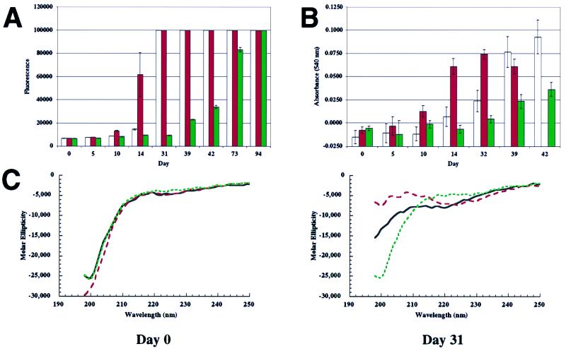
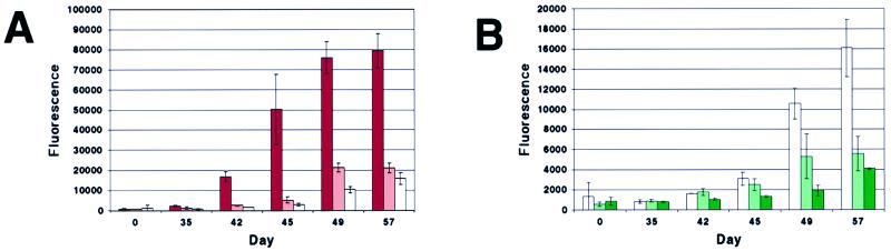
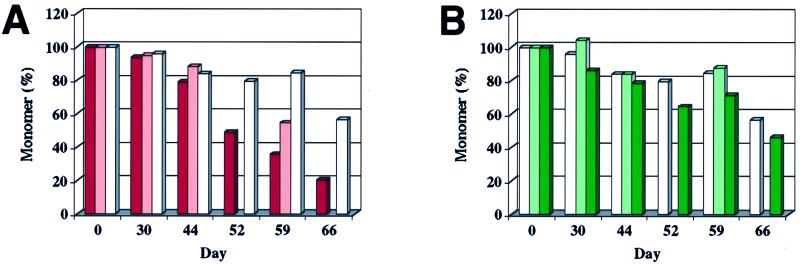
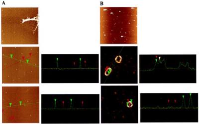
The Parkinson's disease (PD) substantia nigra is characterized by the presence of Lewy bodies containing fibrillar alpha-synuclein. Early-onset PD has been linked to two point mutations in the gene that encodes alpha-synuclein, suggesting that disease may arise from accelerated fibrillization. However, the identity of the pathogenic species and its relationship to the alpha-synuclein fibril has not been elucidated. In this in vitro study, the rates of disappearance of monomeric alpha-synuclein and appearance of fibrillar alpha-synuclein were compared for the wild-type (WT) and two mutant proteins, as well as equimolar mixtures that may model the heterozygous PD patients. Whereas one of the mutant proteins (A53T) and an equimolar mixture of A53T and WT fibrillized more rapidly than WT alpha-synuclein, the other (A30P) and the corresponding equimolar mixture with WT fibrillized more slowly. However, under conditions that ultimately produced fibrils, the A30P monomer was consumed at a comparable rate or slightly more rapidly than the WT monomer, whereas A53T was consumed even more rapidly. The difference between these trends suggested the existence of nonfibrillar alpha-synuclein oligomers, some of which were separated from fibrillar and monomeric alpha-synuclein by sedimentation followed by gel-filtration chromatography. Spheres (range of heights: 2-6 nm), chains of spheres (protofibrils), and rings resembling circularized protofibrils (height: ca. 4 nm) were distinguished from fibrils (height: ca. 8 nm) by atomic force microscopy. Importantly, drug candidates that inhibit alpha-synuclein fibrillization but do not block its oligomerization could mimic the A30P mutation and thus may accelerate disease progression.


Figures

Figure 1
Figure 1
Fibrillization and β-sheet formation of WT, A53T, and A30P α-synuclein (300 μM), followed by three complementary methods. (A) Thio T fluorescence assay shows that A53T fibrillizes most rapidly (red), followed by WT (white) and then A30P (green); signal of 100,000 is maximum (refer to Materials and Methods). (B) Congo red-binding assay also shows that A53T fibrillizes most rapidly (red), followed by WT (white) and then A30P (green). (C) CD spectroscopy detects the random-coil (monomer)-to-β-sheet (fibril) transition. At day 0, all three variants are predominantly random coil. At day 31, the transition to β-sheet structure is most complete in the case of A53T (red), less in the case of WT (black), and has not begun in the case of A30P (green).
Figure 2
Figure 2
Fibrillization (Thio T fluorescence assay) of the three α-synuclein variants, WT, A53T, and A30P (200 μM), and relevant A53T/WT and A30P/WT equimolar mixtures (200 μM total protein). (A) Fibrillization of (left to right) A53T (red), 1:1 A53T/WT (pink), and WT (white). (B) Fibrillization of (left to right) WT (white), 1:1 A30P/WT (light green), and A30P (green). Note expanded y axis relative to A (identical WT run is plotted in both A and B).
Figure 3
Figure 3
Consumption of natively unfolded α-synuclein during fibrillization. Aliquots were removed from incubations run in parallel to those shown in Fig. 2. Quantitation is based on gel-filtration peak weight, but peak height data were comparable. (A) Consumption of A53T (red), 1:1 A53T/WT (pink), and WT (white) during fibrillization (parallels Fig. 2A). (B) Consumption of WT (white), 1:1 A30P/WT (light green), and A30P (green) during fibrillization (parallels Fig. 2B). Where bars are missing, no aliquot was analyzed.
Figure 4
Figure 4
AFM images (and cross-sections) of oligomeric species from 200 μM α-synuclein incubations. (A Top) WT incubation (before sedimentation and gel filtration) showing fibrils and protofibril “spheres” (5-μm square). (Middle) 1:1 A53T/WT incubation after gel filtration showing “spheres” of 5.2–5.5 nm (green cursor and cross-section) and 2.6–3.6 nm (red cursor and cross-section) (2-μm square). (Bottom) 1:1 A30P/WT incubation after gel filtration showing spheres of 4.8–4.9 nm (green cursor and cross-section) and 3.3–3.5 nm (red cursor and cross-section) (2-μm square). (B Top) 1:1 A53T/WT (see A Middle), after incubation, showing rings (5-μm square; very bright features may be amorphous aggregates, with tails from AFM tip “skipping”). (Middle) Close-up of same sample (400-nm square) showing two ring types: circle and ellipse. The periodicity along the ellipse surface is shown in the cross-section to be regular (23 nm), with the maximum height of ca. 4 nm. (Bottom) Another circular ring in the same incubation (300-nm square), showing the diameter of ca. 50 nm and the difference between the maximum (3.6–4.1 nm) and minimum (2.1–2.2 nm) heights.

References

    1. Dunnett S B, Bjorklund A. Nat Suppl. 1999;399:A32–A39. - PubMed
    1. Baba M, Nakajo S, Pang-Hsien T, Tomita T, Nakaya K, Lee V M-Y, Trojanowski J Q, Iwatsubo T. Am J Pathol. 1998;152:879–884. - PMC - PubMed
    1. Spillantini M G, Crowther R A, Jakes R, Hasegawa M, Goedert M. Proc Natl Acad Sci USA. 1998;95:6469–6473. - PMC - PubMed
    1. Selkoe D. Nat Suppl. 1999;399:A23–A31. - PubMed
    1. Kosaka K, Iseki E. Curr Opin Neurol. 1996;9:271–275. - PubMed

Publication types