. 2000 Jan 18;97(2):668-73.
doi: 10.1073/pnas.97.2.668.

The putative tumor suppressors EXT1 and EXT2 form a stable complex that accumulates in the Golgi apparatus and catalyzes the synthesis of heparan sulfate

Affiliations

The putative tumor suppressors EXT1 and EXT2 form a stable complex that accumulates in the Golgi apparatus and catalyzes the synthesis of heparan sulfate

C McCormick et al. Proc Natl Acad Sci U S A. .

Abstract

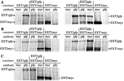
Hereditary multiple exostoses, a dominantly inherited genetic disorder characterized by multiple cartilaginous tumors, is caused by mutations in members of the EXT gene family, EXT1 or EXT2. The proteins encoded by these genes, EXT1 and EXT2, are endoplasmic reticulum-localized type II transmembrane glycoproteins that possess or are tightly associated with glycosyltransferase activities involved in the polymerization of heparan sulfate. Here, by testing a cell line with a specific defect in EXT1 in in vivo and in vitro assays, we show that EXT2 does not harbor significant glycosyltransferase activity in the absence of EXT1. Instead, it appears that EXT1 and EXT2 form a hetero-oligomeric complex in vivo that leads to the accumulation of both proteins in the Golgi apparatus. Remarkably, the Golgi-localized EXT1/EXT2 complex possesses substantially higher glycosyltransferase activity than EXT1 or EXT2 alone, which suggests that the complex represents the biologically relevant form of the enzyme(s). These findings provide a rationale to explain how inherited mutations in either of the two EXT genes can cause loss of activity, resulting in hereditary multiple exostoses.


Figures

Figure 1
Figure 1
Sequence and rescue of the EXT1 defect in sog9 cells. (A) The coding region of murine EXT1 cDNA was amplified from wild-type L cells and HS-deficient sog9 cells by reverse transcription-PCR and analyzed on a 0.8% agarose gel. (B) Sequence analysis indicates that sog9 cells contain a mutation that results in splicing of exon 1 to exon 5 in a +1 reading frame, predicting a truncated 335-aa EXT1 protein. (C) sog9 cells were transfected with the indicated constructs and infected with HSV-1 to detect the presence of newly synthesized cell surface HS. Infected cells stain blue in the presence of 5-bromo-4-chloro-3-indolyl β-d-galactoside (X-Gal).
Figure 2
Figure 2
EXT1 and EXT2 comigrate to the Golgi apparatus. Monolayers of BHK cells were transfected with EXT1-GFP (A), mEXT2-GFP (B), or both (C). When transfected into the same cell, EXT1-GFP and EXT2-GFP relocated to the Golgi (D), while the Golgi apparatus was immunolabeled with an anti-Golgi 58K monoclonal antibody and a Texas red-conjugated secondary antibody (E). When overlaid, they show excellent colocalization (yellow) (F). GFP fusion constructs of the EXT homologs EXTL2 (G) and EXTL3 (H) were also localized, as well as the murine N-deacetylase/N-sulfotransferase (NDST2), a key enzyme in HS biosynthesis (I).
Figure 3
Figure 3
EXT1 and EXT2 form homo- and hetero-oligomers in vivo. BHK cells were transfected with various combinations of Myc-His- or GFP-tagged EXT1 and EXT2 constructs, radiolabeled with [35S]methionine and immunoprecipitated with the indicated anti-Myc or anti-GFP antibodies. (A) EXT1 and EXT2 homo-oligomer analysis. (B) EXT1/EXT2 hetero-oligomer analysis. (C) To determine whether EXT1/EXT2 complex formation was able to occur outside of the cell, lysates containing EXT1-GFP or EXT2-Myc-His were mixed ex vivo and immunoprecipitated with either anti-GFP or anti-Myc antibody.
Figure 4
Figure 4
Mutant EXT1 proteins have different intracellular localizations. Monolayers of BHK cells were tranfected with 335-aa-EXT1-GFP (A), 335–EXT1-GFP and mEXT2-Myc-His (B–D), G339DEXT1-GFP (E), or G339DEXT1-Myc-His and mEXT2-GFP (F–H). After 30 h of expression, Myc-His-tagged EXT1 or EXT2 proteins were detected with an anti-His monoclonal antibody and a Texas red-conjugated secondary antibody.

References

    1. Solomon L. J Bone Joint Surg. 1963;45:292–304.
    1. Leone N C, Shupe J L, Gardner E J, Millar E A, Olson A E, Phillips E C. J Hered. 1987;78:171–177. - PubMed
    1. Hennekam R C. J Med Genet. 1991;28:262–266. - PMC - PubMed
    1. Schmale G A, Conrad E U, Raskind W H. J Bone Joint Surg. 1994;76:986–992. - PubMed
    1. Luckert-Wicklund C, Pauli R, Johnston D, Hecht J. Am J Med Genet. 1995;55:43–46. - PubMed

Publication types

MeSH terms