. 2000 Jan 18;97(2):745-50.
doi: 10.1073/pnas.97.2.745.

Enzymatic reduction of disulfide bonds in lysosomes: characterization of a gamma-interferon-inducible lysosomal thiol reductase (GILT)

Affiliations

Enzymatic reduction of disulfide bonds in lysosomes: characterization of a gamma-interferon-inducible lysosomal thiol reductase (GILT)

B Arunachalam et al. Proc Natl Acad Sci U S A. .

Abstract

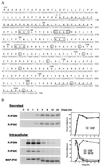
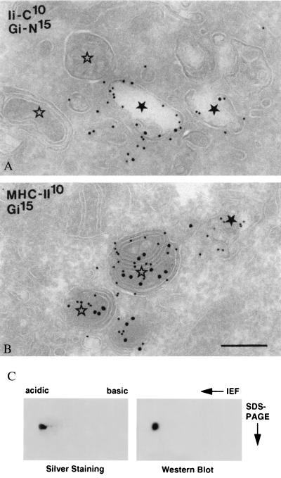
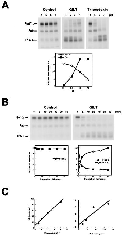
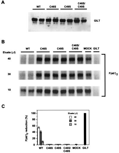
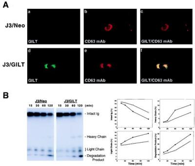
Proteins internalized into the endocytic pathway are usually degraded. Efficient proteolysis requires denaturation, induced by acidic conditions within lysosomes, and reduction of inter- and intrachain disulfide bonds. Cytosolic reduction is mediated enzymatically by thioredoxin, but the mechanism of lysosomal reduction is unknown. We describe here a lysosomal thiol reductase optimally active at low pH and capable of catalyzing disulfide bond reduction both in vivo and in vitro. The active site, determined by mutagenesis, consists of a pair of cysteine residues separated by two amino acids, similar to other enzymes of the thioredoxin family. The enzyme is a soluble glycoprotein that is synthesized as a precursor. After delivery into the endosomal/lysosomal system by the mannose 6-phosphate receptor, N- and C-terminal prosequences are removed. The enzyme is expressed constitutively in antigen-presenting cells and induced by IFN-gamma in other cell types, suggesting a potentially important role in antigen processing.


Figures

Figure 1
Figure 1
(A) GILT cDNA and predicted amino acid sequence. The predicted amino acid sequence of GILT is shown in bold above its nucleotide sequence (GenBank accession no: AF097362). The signal peptide sequence is italicized, propeptide sequences are underlined, potential glycosylation sites are boxed, and cysteines are circled. The active site (CXXC) is enclosed by an oval. (B) Secretion and intracellular processing of GILT. Pala cells were labeled with [35S]methionine for 1 hr and chased for different periods of time. Secreted GILT was precipitated from supernatants with R.IP30N or R.IP30C sera. Intracellular GILT was precipitated from cell extracts with R.IP30N, R.IP30C, or MAP.IP30 antibodies. Samples were analyzed by SDS/PAGE (12%) under reducing conditions and visualized by autoradiography. (Left). The intensity of specific bands is plotted as relative intensity (PD, pixel density) vs. time (hr) (Right). In the lower panel the filled squares represent mature GILT.
Figure 2
Figure 2
Intracellular localization of GILT. Ultrathin cryosections of Raji cells double immunolabeled for the C terminus of invariant chain [rabbit anti-human invariant chain C terminus (IiC)] and the proform of GILT (Gi-N) (A), and MHC class II and mature GILT (Gi) (B). Gold sizes in nanometers are indicated on the plates. In A, IiC and proGILT colocalize in early endosomes ★, whereas MIICs ⋆ are negative. In B, MHC class II is present both in an early endosome ★ and in MIICs ⋆. Mature GILT can be seen only in MIICs. Bars (A and B) = 200 nm. (C) GILT is derivatized with M6P. Affinity-purified mature GILT was separated on isoelectrofocusing followed by SDS/PAGE under reducing conditions. Separated proteins were visualized by silver staining (Left) or transferred to Immobilon P membrane and probed with biotinylated M6PR followed by avidin coupled to horseradish peroxidase (Right).
Figure 3
Figure 3
GILT is an acid optimal thiol reductase. (A) Lysozyme (negative control), GILT (550 nM), or thioredoxin (1.04 μM) was incubated with 125I-F(ab′)2 (55 nM) under different pH conditions for 45 min at 37°C. (B) Lysozyme or GILT (550 nM) was incubated with 125I-F(ab′)2 (55 nM) at pH 4.0 and 37°C, and the reaction was stopped with excess iodoacetamide at different periods of time. (C) To quantitate the effect of substrate denaturation on GILT activity, 125 pM to 1 nM SDS-denatured 125I-F(ab′)2 (Left) and 14.5 nM to 116 nM native 125I-F(ab′)2 (Right) were incubated for 10 min at 37°C with 69 nM or 1.1 μM GILT, respectively, stopping the reaction by the addition of iodoacetamide. Samples were separated by nonreducing SDS/PAGE (12%) and visualized by autoradiography. In A and B, the gels are shown, and the positions of F(ab′)2, Fab, H′ and light chains are indicated (Left). The intensity of specific bands (pixel density) was quantitated and plotted in A as percent reduced heavy and light chains after background subtraction and in B as percent of maximum. In C, the bands were converted to molarity of substrate. The results are presented as Lineweaver–Burke plots.
Figure 4
Figure 4
Identification of the GILT active site by mutagenesis. Wild-type GILT and mutants with Cys-46, Cys-49, or both converted to serine residues were expressed in duplicate in COS-7 cells, immunoisolated, and assayed for thiol reductase activity. GILT and all variants were equally well expressed as determined by Western blotting with rabbit anti-GILT serum (A). B shows the level of reduction of F(ab′)2 by three different concentrations of wild-type and mutant GILT eluates from duplicate transfections, by eluates of various dilutions from a mock transfection, and by purified GILT (0.5 μg). C quantitates the level of reduction by the different eluate concentrations.
Figure 5
Figure 5
Lysosomal reduction in GILT-expressing cells. (A) Immunofluorescence detection of internalized mAb to CD63 (H5C6) in J3/Neo or J3/GILT cells was performed as described (22). After internalization, cells were fixed, permeabilized, and stained for internalized mAb and for GILT (rabbit anti-GILT serum) by using appropriate conjugated secondary antibodies. The internalized mAb colocalizes with GILT. (B) The reduction status of internalized 125I-H5C6 (CD63 mAb) in J3/Neo or J3/GILT transfectants at different periods of time was analyzed by nonreducing SDS/PAGE. (C) Individual bands were quantitated as described and plotted as percent of total in each lane vs. time (min). Open squares represent J3/Neo and closed circles, J3/GILT.

References

    1. Lundstrom-Ljung J, Holmgren A. In: Prolyl Hydroxylase, Protein Disulfide Isomerase, and Other Structurally Related Proteins. Guzman N A, editor. New York: Dekker; 1998. pp. 297–314.
    1. Bardwell J C A, Beckwith J. Cell. 1993;74:769–771. - PubMed
    1. Freedman R B. Curr Opin Struct Biol. 1995;5:85–91. - PubMed
    1. Gilbert H F. In: Prolyl Hydroxylase, Protein Disulfide Isomerase, and Other Structurally Related Proteins. Guzman N A, editor. New York: Dekker; 1998. pp. 341–367.
    1. Holmgren A. Annu Rev Biochem. 1985;54:237–271. - PubMed

Publication types

MeSH terms

Associated data