. 2000 Jan 18;97(2):835-40.
doi: 10.1073/pnas.97.2.835.

Extracellular protein kinase A as a cancer biomarker: its expression by tumor cells and reversal by a myristate-lacking Calpha and RIIbeta subunit overexpression

Affiliations

Extracellular protein kinase A as a cancer biomarker: its expression by tumor cells and reversal by a myristate-lacking Calpha and RIIbeta subunit overexpression

Y S Cho et al. Proc Natl Acad Sci U S A. .

Abstract

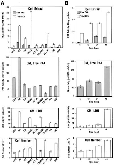
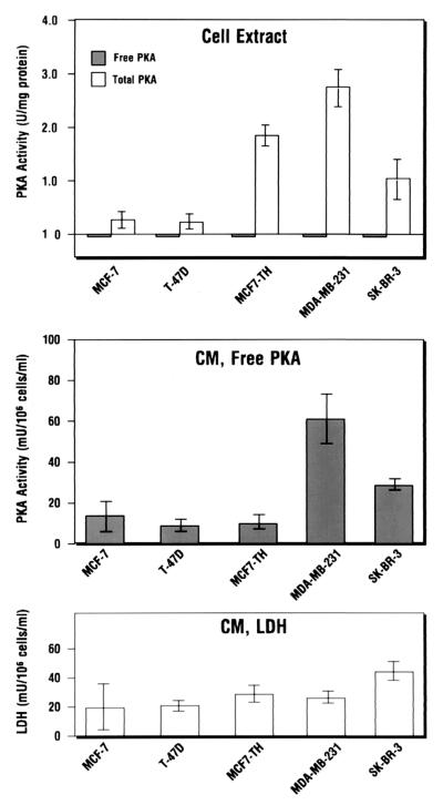
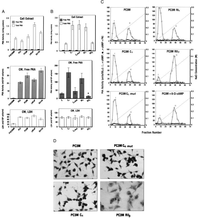
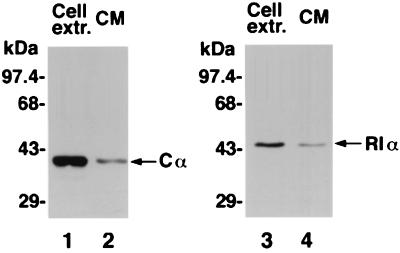
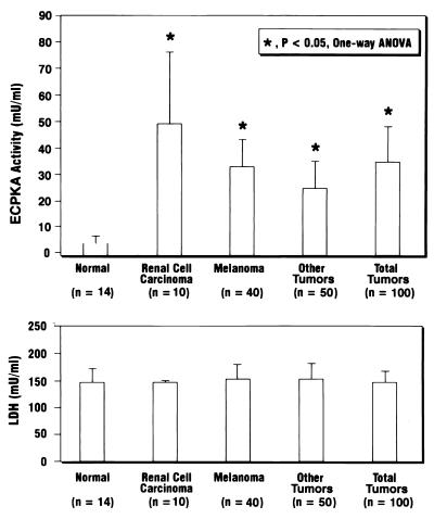
Overexpression of cAMP-dependent protein kinase (PKA) type I isozyme is associated with cell proliferation and neoplastic transformation. The presence of PKA on the external surface of LS-174T human colon carcinoma cells has been shown. Here, we show that cancer cells of various cell types excrete PKA into the conditioned medium. This extracellular PKA (ECPKA) is present in active, free catalytic subunit (C subunit) form, and its activity is specifically inhibited by PKA inhibitory protein, PKI. Overexpression of the Calpha or RIalpha subunit gene of PKA in an expression vector, which up-regulates intracellular PKA type I, markedly up-regulates ECPKA expression. In contrast, overexpression of the RIIbeta subunit, which eliminates PKA type I, up-regulates PKA type II, and reverts the transformed phenotype, down-regulates ECPKA. A mutation in the Calpha gene that prevents myristylation allows the intracellular PKA up-regulation but blocks the ECPKA increase, suggesting that the NH(2)-terminal myristyl group of Calpha is required for the ECPKA expression. In serum of cancer patients, the ECPKA expression is up-regulated 10-fold as compared with normal serum. These results indicate that the ECPKA expression is an ordered cellular response of a living cell to actively exclude excess intracellular PKA molecules from the cell. This phenomenon is up-regulated in tumor cells and has an inverse relationship with the hormone dependency of breast cancer. Thus, the extracellular PKA may serve as a potential diagnostic and prognostic marker for cancer.


Figures

Figure 1
Figure 1
Extracellular protein kinase A expression in the conditioned medium of cancer cells of various cell types. (A) ECPKA expression of lung (A549), bladder (J82, T24, UM-UC-3), colon (HCT-15, CoLo205, LS-174T), and renal (293, 293T) carcinoma cells. (B) Time course of ECPKA expression of T24 bladder carcinomas cells. ■, Free PKA, PKA activity measured in (−) cAMP; □, Total PKA, PKA activity measured in (+) cAMP; Background kinase activity, kinase activity measured in (±) cAMP and (+) PKI. The background activity was subtracted from both free PKA and total PKA; CM, conditioned medium; LDH, Lactate dehydrogenase. For cell extract PKA, both free and total PKA activities are shown in “U/mg protein”; For conditioned medium PKA, only free activity is shown in “mU/106 cells/ml” in 24 hr of culture that contained 2 ml of medium. Cell number, cell count at the end of cell culture (A) or at indicated times (B). Statistical analysis of difference between mean values was performed by the use of a two-tailed t test (statview statistical software; Abacus Concepts, Berkeley, CA).
Figure 2
Figure 2
Extracellular PKA expression is inversely related to hormone dependency of breast cancer cells. PKA activity was measured in cell extracts and conditioned medium of hormone-dependent (MCF-7, T-47D, MCF-7TH) and -independent (SK-BR-3, MDA-MB-231) human breast carcinoma cells by the method described in Experimental Procedures. Statistical analysis was performed by the method described in the Fig. 1 legend. Data represent mean values ± SD of three separate experiments.
Figure 3
Figure 3
Extracellular PKA expression in prostate cancer cells is unrelated to PSA expression and is regulated by intracellular PKA. (A) PKA activity in cell extracts and conditioned medium of prostate cancer cells (PC3, PC3M, LNCap, DU145) and immortalized prostate epithelial cells (PrEC5500). (B) Effect of R and C subunit gene overexpression on intracellular PKA and ECPKA. C, Nontransfected parental cells; Cα, Cα mut, RIα, and RIIβ cells transfected with Cα, Cα mut, RIα, or RIIβ gene in retroviral vector MT-1 (see Experimental Procedures), respectively. PKA and LDH assays were performed as described in Experimental Procedures. Statistical analyses were carried out as described in the legend to Fig. 1. Data in A and B represent mean values ± SD obtained from three separate experiments (*, P < 0.05, vs. that of parental nontransfectant, C). (C) DEAE-column profile of PKA in cells overexpressing PKA subunit genes and cells treated with 8-Cl-cAMP (5 μM for 3 days). PC3M, Parental nontransfectant. For transfectants, see B. Cell extract preparation and DEAE-column chromatography were performed as described in Experimental Procedures. Peak I, PKA-I; Peak II, PKA-II. (D) Effect of PKA subunit overexpression on cell morphology. To examine whole-cell morphology, cells were washed with PBS, fixed with 70% methanol for 5 min, and stained with Giemsa (Sigma) for 15 min. After staining, the whole cells were visualized under an inverted microscope. (Magnification, ×320.)
Figure 4
Figure 4
Immunological characterization of extracellular PKA. Lanes 1 and 2, Western blotting of cell extract and conditioned medium (CM) with anti-Cα antibody. Lanes 3 and 4, Western blotting of cell extract and CM with anti-RIα antibody.
Figure 5
Figure 5
Detection of ECPKA in the serum of cancer patients. Human serum samples were from renal cell carcinoma patients (n = 10); melanoma patients (n = 40); other cancer patients with colon, rectal, adrenal, and lung carcinomas and lymphomas (n = 50); and normal people (n = 14). The statistical analysis of the data was performed by the use of one-way ANOVA (*, P < 0.05). The PKA assay was carried out by the method described in Experimental Procedures with 10 μl of serum samples in the assay (total volume, 50 μl). LDH assay was carried out as described in Experimental Procedures with 10 μl of 6-fold-diluted serum.

References

    1. Krebs E G, Beavo J A. Annu Rev Biochem. 1979;48:923–939. - PubMed
    1. Beebe S J, Corbin J D. The Enzymes: Control by Phosphorylation. Vol. 17. New York: Academic; 1986. pp. 43–111.
    1. McKnight G S, Clegg C H, Uhler M D, Chrivia J C, Cadd G G, Correll L A, Otten A D. Recent Prog Horm Res. 1988;44:307–335. - PubMed
    1. Levy F O, Oyen O, Sandberg M, Tasken K, Eskild W, Hansson V, Jahnsen T. Mol Endocrinol. 1988;2:1364–1373. - PubMed
    1. Lohmann S M, Walter U. In: Advances in Cyclic Nucleotide and Protein Phosphorylation Research. Greengard P, Robison G A, editors. Vol. 18. New York: Raven; 1984. pp. 63–117. - PubMed

MeSH terms

LinkOut - more resources