. 2000 Jan 18;97(2):841-6.
doi: 10.1073/pnas.97.2.841.

Aortic wall damage in mice unable to synthesize ascorbic acid

Affiliations

Aortic wall damage in mice unable to synthesize ascorbic acid

N Maeda et al. Proc Natl Acad Sci U S A. .

Abstract

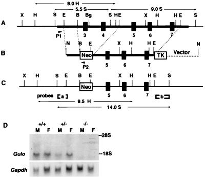
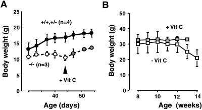
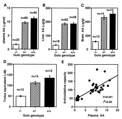
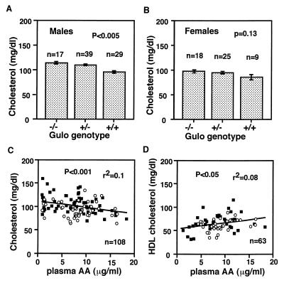
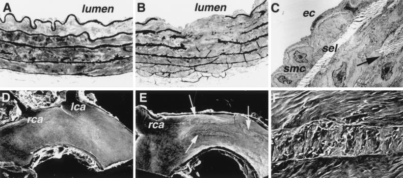
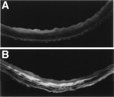
By inactivating the gene for L-gulono-gamma-lactone oxidase, a key enzyme in ascorbic acid synthesis, we have generated mice that, like humans, depend on dietary vitamin C. Regular chow, containing about 110 mg/kg of vitamin C, is unable to support the growth of the mutant mice, which require L-ascorbic acid supplemented in their drinking water (330 mg/liter). Upon withdrawal of supplementation, plasma and tissue ascorbic acid levels decreased to 10-15% of normal within 2 weeks, and after 5 weeks the mutants became anemic, began to lose weight, and die. Plasma total antioxidative capacities were approximately 37% normal in homozygotes after feeding the unsupplemented diet for 3-5 weeks. As plasma ascorbic acid decreased, small, but significant, increases in total cholesterol and decreases in high density lipoprotein cholesterol were observed. The most striking effects of the marginal dietary vitamin C were alterations in the wall of aorta, evidenced by the disruption of elastic laminae, smooth muscle cell proliferation, and focal endothelial desquamation of the luminal surface. Thus, marginal vitamin C deficiency affects the vascular integrity of mice unable to synthesize ascorbic acid, with potentially profound effects on the pathogenesis of vascular diseases. Breeding the vitamin C-dependent mice with mice carrying defined genetic mutations will provide numerous opportunities for systematic studies of the role of antioxidants in health and disease.


Figures

Figure 1
Figure 1
Targeted modification of the mouse Gulo gene. (A) The endogenous mouse Gulo locus. The thick horizontal bar indicates the phage clone containing exons 3–7, marked by black boxes. (B) The targeting vector. Arrows P1 and P2 are relative positions of primers used to screen embryonic stem cells for the targeting event. (C) The inactivated Gulo locus. [a] and [b] are probes used for Southern blottings. Sizes and restriction enzymes of fragments diagnostic for the modification are indicated. B, BamHI; Bg, BglII; E, EcoRI; H, HindIII; N, NotI; S, SacI; X, XbaI. (D) Northern blot analysis of the liver RNA (20 mg) isolated from the livers of a male and a female of the three Gulo genotypes. The filter was hybridized with a Gulo probe (Upper), washed, and rehybridized with a glyceraldehyde-3-phosphate dehydrogenase gene (Gapdh) probe (Lower). The positions of 28S and 18S RNA are marked.
Figure 2
Figure 2
Body weight changes of Gulo −/− mice. (A) Females from a single litter from a heterozygous pair weaned on regular chow at age 21 days. Mean weights ± SD of one wild type plus three heterozygotes (●) and of three homozygotes (○) are shown. At day 45, vitamin C (330 mg/liter) in the drinking water was given to the animals (stippled circle). Because one homozygote died at day 47, the values at days 51 and 54 are the average weights of two homozygotes. Differences between two groups were statistically significant at all time points. (B) Nine Gulo −/− males were raised on chow with vitamin C supplementation to 8 weeks of age and then supplementation was withdrawn from six of the mice. Mean body weights ± SD for three mice with vitamin C supplementation (stippled square) and for six mice without supplementation (□) are shown, except that the 14th week was for three mice. P < 0.01 at 13-week time point.
Figure 3
Figure 3
Ascorbic acid (AA) levels in plasma (A), liver (B), and brain (C). Homozygous (−/−) mice were on chow diet without vitamin C supplement for 3–5 weeks. Data are expressed mean ± SE. P < 0.0001. (D) Plasma total antioxidative capacity expressed as trolox equivalent (μM). P < 0.0001. (E) Correlation between plasma ascorbic acid and total antioxidative capacity of individual animals including all three genotypes.
Figure 4
Figure 4
Plasma cholesterol levels in the Gulo mutant males (A) and females (B). Mean ± SEM. Homozygotes (−/−) were on chow with vitamin C supplement until 3–5 weeks before the determinations. Heterozygotes and wild-type mice were on unsupplemented chow at all times. Correlation between plasma ascorbic acid (AA) and plasma total cholesterol (C) and HDL cholesterol (D) levels in individual animals including all three genotypes. Some homozygotes were with vitamin C supplement and others were unsupplemented. ○, females; ■, males.
Figure 5
Figure 5
Abnormalities in the aorta of Gulo −/− mice. (A) A cross section of the descending thoracic aorta from a wild-type mouse showing uninterrupted elastic laminae. Toluidine blue staining, light microscopy, ×400. (B) A cross section of the descending thoracic aorta from a Gulo −/− mouse with disrupted elastic laminae. Light microscopy, ×400. (C) TEM of the area adjacent to that shown in B. A single layer of mildly activated smooth muscle cells (smc) is present in the intima between endothelial cells (ec) and superficial elastic lamina (sel). A break in the elastica is marked by an arrow. Magnification: ×4,000. (D) Scanning electron microscopy of the aortic arch of a wild-type mouse showing a normal luminal surface. lca, left carotid artery; rca, right carotid artery. Magnification: ×20. (E) Scanning electron microscopy of the aortic arch of an Gulo −/− mouse showing multiple longitudinal surface defects (arrows). Magnification: ×20. (F) Higher magnification (×200) of the defect.
Figure 6
Figure 6
Evans blue extravasation in the ascending aorta of (A) a wild-type mouse and (B) a Gulo −/− mouse. Magnification: ×200. Image processing was used to subtract green autofluorescence, and the brighter area in this black and white figure corresponds to the red fluorescence of Evans blue.

References

    1. Knight J A. Ann Clin Lab Sci. 1995;25:111–121. - PubMed
    1. Berliner J A, Heinecke J W. Free Radical Biol Med. 1996;20:707–727. - PubMed
    1. Collins A R. BioEssays. 1999;21:238–246. - PubMed
    1. Bowling A C, Beal M F. Life Sci. 1995;56:1151–1171. - PubMed
    1. Smithies O, Maeda N. Proc Natl Acad Sci USA. 1995;92:5266–5272. - PMC - PubMed

Publication types

MeSH terms