. 2000 Feb;68(2):621-9.
doi: 10.1128/IAI.68.2.621-629.2000.

CD4(+) T-cell subsets that mediate immunological memory to Mycobacterium tuberculosis infection in mice

Affiliations

CD4(+) T-cell subsets that mediate immunological memory to Mycobacterium tuberculosis infection in mice

P Andersen et al. Infect Immun. 2000 Feb.

Abstract

We have studied CD4(+) T cells that mediate immunological memory to an intravenous infection with Mycobacterium tuberculosis. The studies were conducted with a mouse model of memory immunity in which mice are rendered immune by a primary infection followed by antibiotic treatment and rest. Shortly after reinfection, tuberculosis-specific memory cells were recruited from the recirculating pool, leading to rapidly increasing precursor frequencies in the liver and a simultaneous decrease in the blood. A small subset of the infiltrating T cells was rapidly activated (<20 h) and expressed high levels of intracellular gamma interferon and the T-cell activation markers CD69 and CD25. These memory effector T cells expressed intermediate levels of CD45RB and were heterogeneous with regard to the L-selectin and CD44 markers. By adoptive transfer into nude mice, the highest level of resistance to a challenge with M. tuberculosis was mediated by CD45RB(high), L-selectin(high), CD44(low) cells. Taken together, these two lines of evidence support an important role for memory cells which have reverted to a naive phenotype in the long-term protection against M. tuberculosis.


Figures

FIG. 1
FIG. 1
Cellular infiltration and granuloma formation in the livers of naive and memory immune mice challenged with M. tuberculosis. Photomicrographs of livers from naive (top panel) and memory immune (bottom panel) mice at days 2 to 14 of infection with M. tuberculosis are shown. The granulomas are often initially situated in the perivascular regions around portal veins and at later time points are scattered throughout the liver parenchyme. Hematoxylin-eosin staining was used. Magnification, ×300.
FIG. 2
FIG. 2
Recruitment of recirculating M. tuberculosis-specific memory cells to the liver during the course of tuberculosis infection. Lymphocytes were purified from the blood and liver and used for ELISPOT analysis of M. tuberculosis ST-CF-specific precursors. Each point represents the mean and standard error of the mean of data from five individual mice. Results at all timepoints throughout infection were significantly different from those at day zero. The experiment was repeated with similar results.
FIG. 3
FIG. 3
Two-color flow cytometric analysis of cell size and surface marker expression on CD4+ cells isolated from the blood and livers of memory immune mice before (day 0) and during the course of infection with M. tuberculosis. Lymphocytes were pooled from four to six animals for each time point. The experiment was repeated with the same overall results.
FIG. 4
FIG. 4
Definition of CD4+ memory effector T cells by three-color flow cytometric analysis of activation markers (CD69), intracellular cytokines (IFN-γ), and cellular size. Memory immune mice (A) and naive mice (B) were challenged with M. tuberculosis, and at 20 h postinfection liver lymphocytes from four mice were purified and pooled. Gated CD4+ T cells from four mice were analyzed for CD69 and intracellular IFN-γ. Cell size is shown for activated and cytokine-expressing memory effector cells (quadrant 2) versus normal nonactivated T cells (quadrant 3).
FIG. 5
FIG. 5
Three-color flow cytometric analysis of the phenotype of cytokine-producing memory effector T cells. Pooled lymphocytes from the livers and spleens of memory immune mice (n = 5) isolated 20 h after the infection with M. tuberculosis were analyzed for intracellular IFN-γ and the expression of the surface molecules L-selectin, CD44, and CD45RB. The cutoff for positive intracellular IFN-γ was based on anti-IFN-γ isotype controls for each combination. The experiment was repeated with the same results.
FIG. 6
FIG. 6
Isolation of CD4+ CD45RBlow and CD45RBhigh subsets by MACS separation. CD4+ T cells from pooled spleen and blood cells from memory immune mice (n = 12) were sorted and analyzed for CD45RB, L-selectin, and CD44 expression. The subsets purified from naive mice had similar profiles.
FIG. 7
FIG. 7
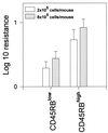
Adoptive protection by 2 × 10⁵ and 8 × 10⁵ purified CD45RBlow and CD45RBhigh cells. The purified subsets were transferred into nude mice which were challenged with M. tuberculosis, and bacteria in the spleen were enumerated 14 days later. Data are given as log10 resistance (for the calculation, see Materials and Methods).
FIG. 8
FIG. 8
Adoptive protection of M. tuberculosis-infected liver, spleen, and lung by purified T-cell subsets. Purified CD4+ T-cell subsets (8 × 10⁵ cells/mouse) from naive and memory immune mice were transferred into nude mice, and bacteria were enumerated 14 days after challenge with M. tuberculosis. Data are given as log10 resistance. Significant differences between protection transferred by cells from naive and memory immune mice are indicated by asterisks.

References

    1. Akbar A N, Terry L, Timms A, Beverley P C, Janossy G. Loss of CD45R and gain of UCHL1 reactivity is a feature of primed T cells. J Immunol. 1988;140:2171–2178. - PubMed
    1. Andersen P, Andersen A B, Sorensen A L, Nagai S. Recall of long-lived immunity to Mycobacterium tuberculosis infection in mice. J Immunol. 1995;154:3359–3372. - PubMed
    1. Andersen P, Askgaard D, Ljungqvist L, Bennedsen J, Heron I. Proteins released from Mycobacterium tuberculosis during growth. Infect Immunol. 1991;59:1905–1910. - PMC - PubMed
    1. Andersen P, Askgaard D, Ljungqvist L, Bentzon M W, Heron I. T-cell proliferative response to antigens secreted by Mycobacterium tuberculosis. Infect Immun. 1991;59:1558–1563. - PMC - PubMed
    1. Andersen P, Heron I. Specificity of a protective memory immune response against Mycobacterium tuberculosis. Infect Immun. 1993;61:844–851. - PMC - PubMed

Publication types