. 2000 Feb;68(2):637-43.
doi: 10.1128/IAI.68.2.637-643.2000.

Streptococcus suis serotype 2 interactions with human brain microvascular endothelial cells

Affiliations

Streptococcus suis serotype 2 interactions with human brain microvascular endothelial cells

N Charland et al. Infect Immun. 2000 Feb.

Abstract

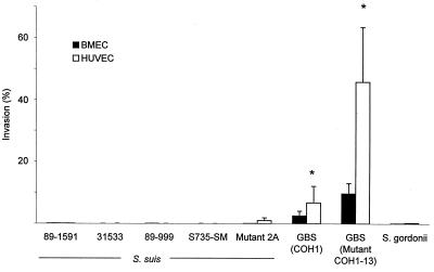
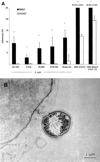
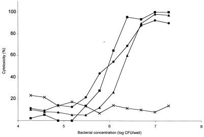
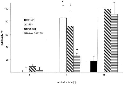
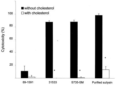
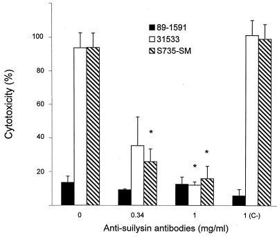
Streptococcus suis serotype 2 is a worldwide causative agent of many forms of swine infection and is also recognized as a zoonotic agent causing human disease, including meningitis. The pathogenesis of S. suis infections is poorly understood. Bacteria circulate in the bloodstream in the nonimmune host until they come in contact with brain microvascular endothelial cells (BMEC) forming the blood-brain barrier. The bacterial polysaccharide capsule confers antiphagocytic properties. It is known that group B streptococci (GBS) invade and damage BMEC, which may be a primary step in the pathogenesis of neonatal meningitis. Interactions between S. suis and human endothelial cells were studied to determine if they differ from those between GBS and endothelial cells. Invasion assays performed with BMEC and human umbilical vein endothelial cells demonstrated that unlike GBS, S. suis serotype 2 could not invade either type of cell. Adherence assays showed that S. suis adhered only to BMEC, whereas GBS adhered to both types of cell. These interactions were not affected by the presence of a capsule, since acapsular mutants from both bacterial species adhered similarly compared to the wild-type strains. Lactate dehydrogenase release measurements indicated that some S. suis strains were highly cytotoxic for BMEC, even more than GBS, whereas others were not toxic at all. Cell damage was related to suilysin (S. suis hemolysin) production, since only suilysin-producing strains were cytotoxic and cytotoxicity could be inhibited by cholesterol and antisuilysin antibodies. It is possible that hemolysin-positive S. suis strains use adherence and suilysin-induced BMEC injury, as opposed to direct cellular invasion, to proceed from the circulation to the central nervous system.


Figures

FIG. 1
FIG. 1
Invasion of BMEC and HUVEC by S. suis 89-1591, 31533, 89-999, and S735-SM and acapsular mutant 2A and GBS strain COH1 and acapsular mutant COH1-13. S. gordonii was used as a noninvasive control. ∗, P < 0.05 versus wild-type strain COH1. Results were determined after a 2-h exposure with 100-μl aliquots of 106 CFU/ml, followed by an additional 2-h incubation in the presence of penicillin-gentamicin to kill extracellular bacteria, and BMEC lysis to retrieve 100-μl aliquots of intracellular bacteria for viable plate counts.
FIG. 2
FIG. 2
(A) Adherence to BMEC and HUVEC by S. suis 89-1591, 31533, 89-999, and S735-SM and acapsular mutant 2A and GBS strain COH1 and acapsular mutant COH1-13. ∗, P < 0.05 versus adherence to HUVEC. Results were determined after a 2-h exposure with 100-μl aliquots of 106 CFU/ml, followed by extensive washing of nonadherent bacteria and BMEC lysis to retrieve 100-μl aliquots of total cell-associated bacteria for viable plate counts. (B) Transmission electron micrograph showing S. suis adherent to BMEC.
FIG. 3
FIG. 3
Effect of S. suis concentration on BMEC injury. Symbols: ●, hemolytic strain 31533; ■, hemolytic strain S735-SM (wild-type strain); ▴, weakly hemolytic mutant C3P2E5; ×: nonhemolytic strain 89-1591. BMEC cytotoxicity was determined by measuring LDH release in the presence of different concentrations of S. suis strains after a 4-h incubation. Results of a representative experiment are shown.
FIG. 4
FIG. 4
Effect of time of incubation with S. suis hemolytic strains 31533 and S735-SM (wild type strain), weakly hemolytic mutant C3P2E5, and nonhemolytic strain 89-1591 on BMEC injury. ∗, P < 0.05 versus 2-h incubation; ∗∗, P < 0.05 versus S735-SM (4 h). BMEC cytotoxicity was determined by measuring LDH release in the presence of 100-μl aliquots of 108 CFU/ml after different incubation times.
FIG. 5
FIG. 5
Effect of increasing concentrations of purified suilysin on BMEC injury. BMEC cytotoxicity was determined by measuring LDH release in the presence of different concentrations of purified suilysin after a 4-h incubation.
FIG. 6
FIG. 6
Inhibition by cholesterol of S. suis cytotoxicity for BMEC. ∗, P < 0.05 versus no inhibitor. We incubated 108 CFU/ml or purified suilysin at 4.6 μg/ml in the absence and presence of 2 mg (bacteria) or 100 μg (suilysin) of cholesterol per ml for 1 h at 37°C before incubation of 100-μl aliquots with BMEC for 4 h. BMEC cytotoxicity was determined by measuring LDH release.
FIG. 7
FIG. 7
Inhibition by antisuilysin antibodies of S. suis cytotoxicity for BMEC. ∗, P < 0.05 versus the no-antibody or nonspecific antilipopolysaccharide antibody (C−) control. We incubated 108 CFU/ml in the presence of increasing antibody concentrations for 1 h at 37°C before incubation of 100-μl aliquots with BMEC for 4 h. BMEC cytotoxicity was determined by measuring LDH release.
FIG. 8
FIG. 8
Transmission electron micrographs demonstrating BMEC injury caused by S. suis at 108 CFU/ml. Panels: A, suilysin-producing strain 31533; B, non-suilysin-producing strain 89-1591; C, no-bacterium control. BMEC integrity after a 2-h incubation with strain 89-1591 (B) was comparable to that of the no-bacterium control (C).

References

    1. Alouf J E, Geoffroy C. The family of the antigenically-related cholesterol-binding (“sulphydryl-activated”) cytolytic toxins. In: Alouf J E, editor. Sourcebook of bacterial protein toxins. New York, N.Y: Academic Press, Inc.; 1991. pp. 147–186.
    1. Arends J P, Zanen H C. Meningitis caused by Streptococcus suis in humans. J Infect Dis. 1988;10:131–137. - PubMed
    1. Baker C J, Edwards M S. Group B streptococcal infections. In: Klein J S, Klein J O, editors. Infectious diseases of the fetus and the newborn. Philadelphia, Pa: The W. B. Saunders Co.; 1990. pp. 742–811.
    1. Betz A L, Goldstein G W. Specialized properties and solute transport in brain capillaries. Annu Rev Physiol. 1986;48:241–250. - PubMed
    1. Boulnois G J, Paton J C, Mitchell T J, Andrew P W. Structure and function of pneumolysin, the multifunctional, thiol-activated toxin of Streptococcus pneumoniae. Mol Microbiol. 1991;5:2611–2616. - PubMed

Publication types

LinkOut - more resources