. 2000 Feb;68(2):708-15.
doi: 10.1128/IAI.68.2.708-715.2000.

Identification of an antigen localized to an apparent septum within dividing chlamydiae

Affiliations

Identification of an antigen localized to an apparent septum within dividing chlamydiae

W J Brown et al. Infect Immun. 2000 Feb.

Abstract

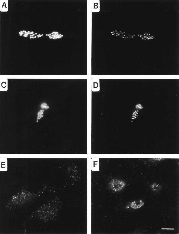
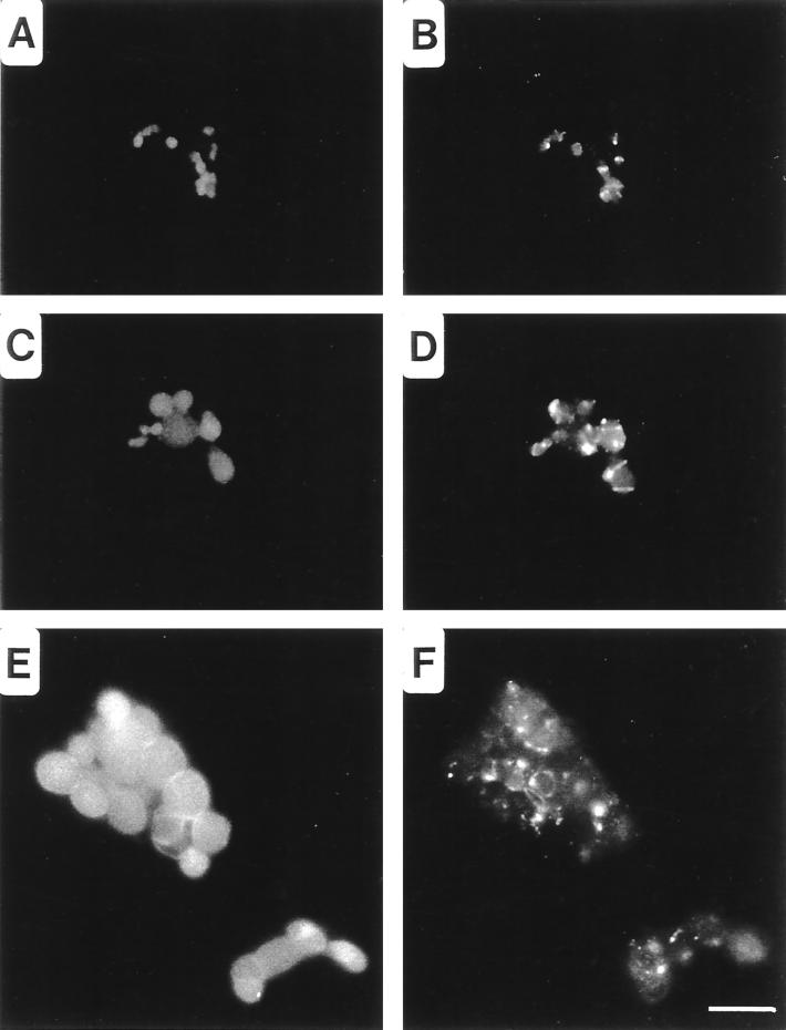
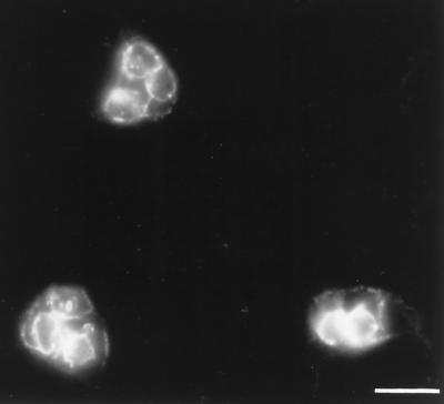
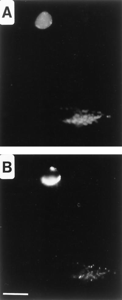
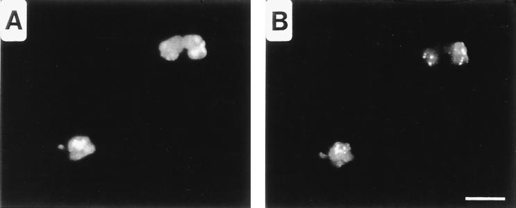
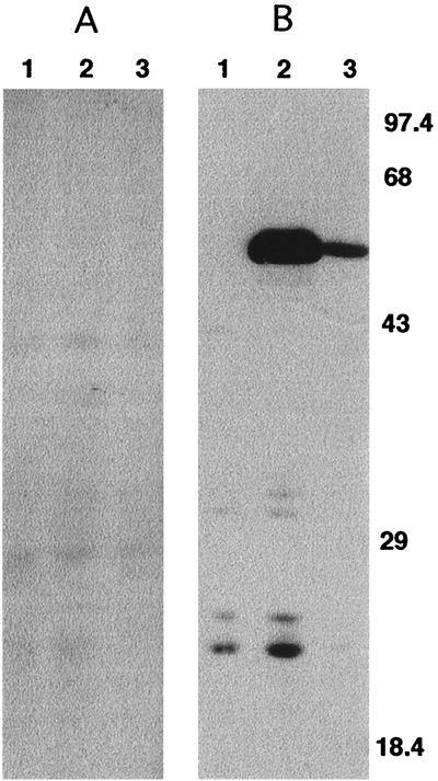
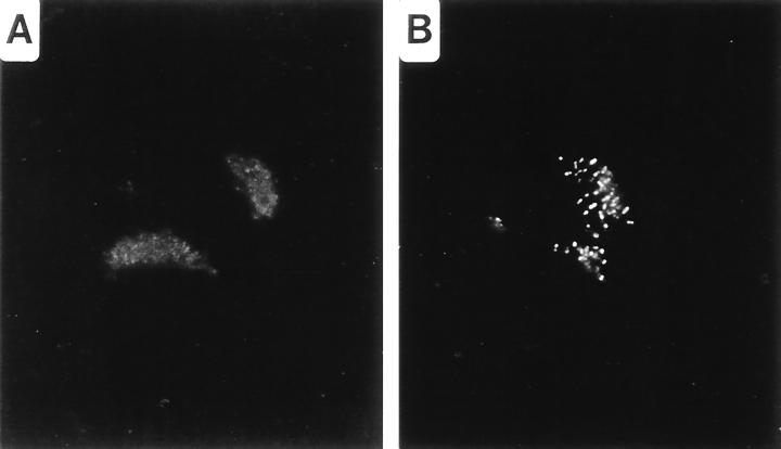
The process of chlamydial cell division has not been thoroughly investigated. The lack of detectable peptidoglycan and the absence of an FtsZ homolog within chlamydiae suggest an unusual mechanism for the division process. Our laboratory has identified an antigen (SEP antigen) localized to a ring-like structure at the apparent septum within dividing chlamydial reticulate bodies (RB). Antisera directed against SEP show similar patterns of antigen distribution in Chlamydia trachomatis and Chlamydia psittaci RB. In contrast to localization in RB, SEP in elementary bodies appears diffuse and irregular, suggesting that the distribution of the antigen is developmental-stage specific. Treatment of chlamydiae with inhibitors of peptidoglycan synthesis or culture of chlamydiae in medium lacking tryptophan leads to the formation of nondividing, aberrant RB. Staining of aberrant RB with anti-SEP reveals a marked redistribution of the antigen. Within C. trachomatis-infected cells, ampicillin treatment leads to high levels of SEP accumulation at the periphery of aberrant RB, while in C. psittaci, treatment causes SEP to localize to distinct punctate sites within the bacteria. Aberrancy produced via tryptophan depletion results in a different pattern of SEP distribution. In either case, the reversal of aberrant formation results in the production of normal RB and a redistribution of SEP to the apparent plane of bacterial division. Collectively these studies identify a unique chlamydial-genus-common and developmental-stage-specific antigen that may be associated with RB division.


Figures

FIG. 1
FIG. 1
Distribution of SEP in C. psittaci- and C. trachomatis-infected HeLa cells visualized by fluorescence microscopy. C. psittaci-infected cells fixed with methanol 18 hpi were doubly labeled with anti-HSP60 (A) and anti-SEP (B) antibodies or with anti-LPS (C) and anti-SEP (D) antibodies. Notice that SEP localizes to the midpoint within dividing RB. SEP distribution is altered in chlamydiae found in late inclusions. (E) SEP distribution within C. psittaci-infected cells fixed 30 hpi. (F) SEP is a genus-common antigen, as is shown by labeling of C. trachomatis-infected cells fixed 18 hpi with anti-SEP antibodies. Note that in these cells the antigen is distributed in both bar and ring structures. Bar, 5 μm.
FIG. 2
FIG. 2
Redistribution of SEP in aberrant C. psittaci RB produced following the addition of ampicillin to the culture medium. Fluorescence microscopy was performed by double-labeling cells with anti-HSP60 (A, C, and E) and anti-SEP (B, D, and F) antibodies. C. psittaci-infected HeLa cells were cultured in MEM-10 for 10 hpi and then cultured in MEM-10 containing 10 μg of ampicillin/ml for an additional 4 (A and B) or 16 (C and D) h prior to methanol fixation. In addition, C. psittaci-infected HeLa cells were treated with 10 μg of ampicillin/ml immediately postinfection for 24 h prior to methanol fixation (E and F). Bar, 5 μm.
FIG. 3
FIG. 3
Redistribution of SEP in aberrant C. trachomatis RB produced following the addition of ampicillin to the culture medium. C. trachomatis-infected HeLa cells were treated with 0.2 μg of ampicillin/ml for 24 h prior to methanol fixation. Fluorescence microscopy was performed by labeling cells with anti-SEP antibodies. Note the greatly enlarged RB and the anti-SEP staining at the peripheries. Bar, 5 μm.
FIG. 4
FIG. 4
Distribution of SEP after removal of MEM-10 containing ampicillin. C. psittaci-infected HeLa cells were cultured in MEM-10 containing 10 μg of ampicillin/ml for 24 h, after which the medium was removed and replaced with MEM-10 lacking ampicillin for 14 h prior to methanol fixation. Fluorescence microscopy was performed by double-labeling cells with anti-HSP60 (A) and anti-SEP (B) antibodies. Bar, 5 μm.
FIG. 5
FIG. 5
Distribution of SEP in aberrant RB of C. psittaci produced by Trp starvation. C. psittaci-infected HeLa cells were cultured in Trp MEM-1 for 46 hpi prior to methanol fixation. Fluorescence microscopy was performed by double-labeling cells with anti-HSP60 (A) and anti-SEP (B) antibodies. Bar, 5 μm.
FIG. 6
FIG. 6
Western blot analysis of SEP and HSP60 from C. trachomatis-infected HeLa cell lysates. Anti-SEP (A) and anti-HSP60 (B) antibodies were used to probe lysates collected from 18-h mock-infected HeLa cells cultured in MEM-10 (lanes 1), 40-h C. trachomatis-infected HeLa cells cultured in MEM-10 containing 0.2 μg of ampicillin/ml (lanes 2), or 18-h C. trachomatis-infected HeLa cells cultured in MEM-10 (lanes 3). Molecular mass standards (in kilodaltons) are shown on the right.
FIG. 7
FIG. 7
Immunoadsorption of MCWS with anti-SEP antibodies. Anti-SEP antibodies were absorbed with purified MCWS (A) or mock absorbed with BSA (B). Absorbed antisera were used to probe 18-h methanol-fixed C. psittaci-infected HeLa cells.

References

    1. Addinall S G, Lutkenhaus J. FtsA is localized to the septum in an FtsZ-dependent manner. J Bacteriol. 1996;178:7167–7172. - PMC - PubMed
    1. Beall B, Lutkenhaus J. FtsZ in Bacillus subtilis is required for vegetative septation and for asymmetric septation during sporulation. Genes Dev. 1991;5:447–455. - PubMed
    1. Beatty W L, Belanger T A, Desai A A, Morrison R P, Byrne G I. Tryptophan depletion as a mechanism of gamma interferon-mediated chlamydial persistence. Infect Immun. 1994;62:3705–3711. - PMC - PubMed
    1. Beatty W L, Byrne G I, Morrison R P. Morphologic and antigenic characterization of interferon-gamma-mediated persistent Chlamydia trachomatis infection in vitro. Proc Natl Acad Sci USA. 1993;90:3998–4002. - PMC - PubMed
    1. Bi E, Lutkenhaus J. FtsZ ring structure associated with division in Escherichia coli. Nature. 1991;354:161–164. - PubMed

Publication types

LinkOut - more resources