. 2000 Feb;68(2):767-78.
doi: 10.1128/IAI.68.2.767-778.2000.

Disruption of the genes encoding antigen 85A and antigen 85B of Mycobacterium tuberculosis H37Rv: effect on growth in culture and in macrophages

Affiliations

Disruption of the genes encoding antigen 85A and antigen 85B of Mycobacterium tuberculosis H37Rv: effect on growth in culture and in macrophages

L Y Armitige et al. Infect Immun. 2000 Feb.

Abstract

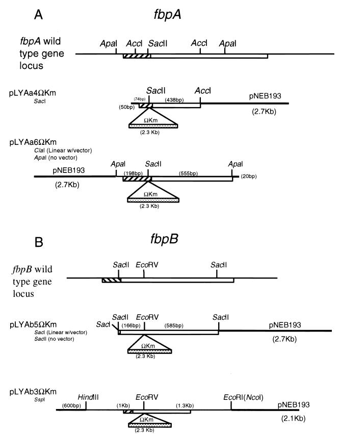
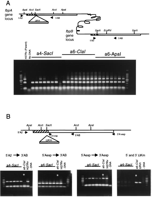
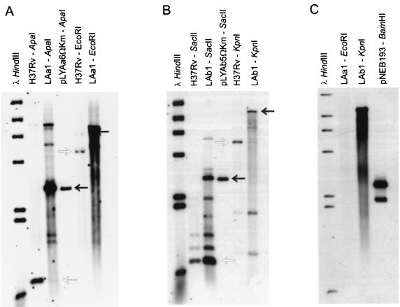
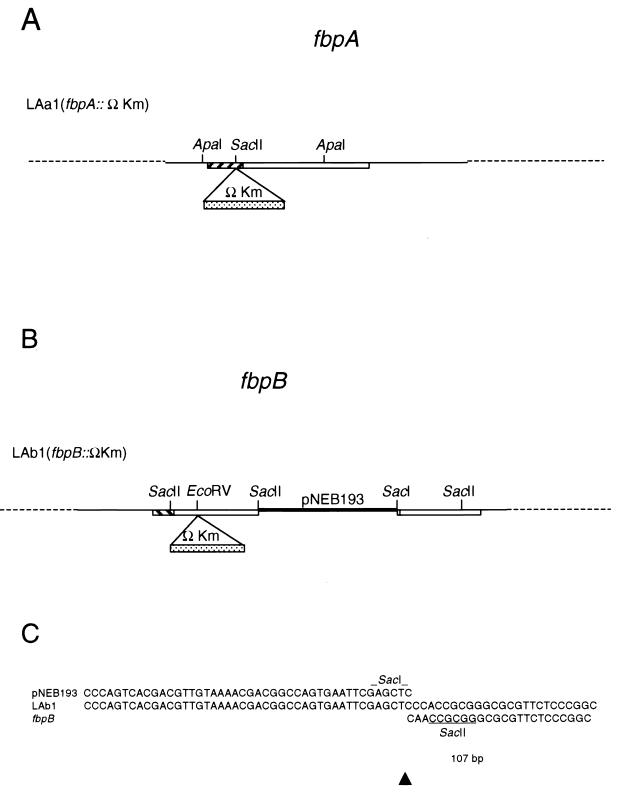
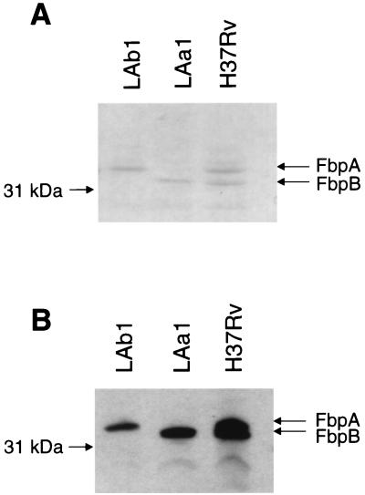
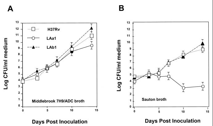
The mechanism of pathogenesis of Mycobacterium tuberculosis is thought to be multifactorial. Among the putative virulence factors is the antigen 85 (Ag85) complex. This family of exported fibronectin-binding proteins consists of members Ag85A, Ag85B, and Ag85C and is most prominently represented by 85A and 85B. These proteins have recently been shown to possess mycolyl transferase activity and likely play a role in cell wall synthesis. The purpose of this study was to generate strains of M. tuberculosis deficient in expression of the principal members of this complex in order to determine their role in the pathogenesis of M. tuberculosis. Constructs of fbpA and fbpB disrupted with the kanamycin resistance marker OmegaKm and containing varying amounts of flanking gene and plasmid vector sequences were then introduced as linear fragments into H37Rv by electroporation. Southern blot and PCR analyses revealed disruption of the homologous gene locus in one fbpA::OmegaKm transformant and one fbpB::OmegaKm transformant. The fbpA::OmegaKm mutant, LAa1, resulted from a double-crossover integration event, whereas the fbpB::OmegaKm variant, LAb1, was the product of a single-crossover type event that resulted in insertion of both OmegaKm and plasmid sequences. Sodium dodecyl sulfate-polyacrylamide gel electrophoresis and Western blot analysis confirmed that expression of the disrupted gene was not detectable in the fbpA and fbpB mutants. Analysis of growth rates demonstrated that the fbpB mutant LAb1 grew at a rate similar to that of the wild-type parent in enriched and nutrient-poor laboratory media as well as in human (THP-1) and mouse (J774.1A) macrophage-like cell lines. The fbpA mutant LAa1 grew similarly to the parent H37Rv in enriched laboratory media but exhibited little or no growth in nutrient-poor media and macrophage-like cell lines. The targeted disruption of two genes encoding mycolyl transferase and fibronectin-binding activities in M. tuberculosis will permit the systematic determination of their roles in the physiology and pathogenesis of this organism.


Figures

FIG. 1
FIG. 1
Plasmids generated for transformation of M. tuberculosis H37Rv. Open boxes, fbpA and fbpB open reading frames; hatched boxes, signal sequences; heavy lines, vector sequences. Numbers in parentheses are distances between restriction sites. (A) fbpA::ΩKm constructs. A 500-bp AccI fragment and a 750-bp ApaI fragment were cloned from fbpA into pNEB193, a ColE1 plasmid, and mutated by the addition of an ΩKm cassette at the SacII site. Plasmid pLYAa4ΩKm was digested with SacI and introduced into H37Rv as a linear fragment, while pLYAa6ΩKm was electroporated into H37Rv associated with (by digestion with ClaI) and liberated from (by digestion with ApaI) the vector pNEB193. (B) fbpB::ΩKm constructs. A 750-bp ApaI fragment and a 2.3-kb HindIII/NcoI fragment were cloned from fbpB into pNEB193 and mutated by the addition of an ΩKm cassette at the EcoRV site. Plasmid pLYAb5ΩKm was introduced into H37Rv associated with (by SacI digestion) and liberated from (by SacII digestion) the vector pNEB193.
FIG. 2
FIG. 2
PCR analysis of M. tuberculosis H37Rv transformed with constructs pLYAa4ΩKm treated with SacI (a4-SacI), pLYAa6ΩKm treated with ClaI (a6-ClaI), and pLYAa6ΩKm treated with ApaI (a6-ApaI). (A) Initial screening strategy. The locations of the primers used are indicated in the diagram. DNA from pLYAa4ΩKm- and pLYAa6ΩKm-transformed Kmr colonies was used as a template in a PCR using three primers: one unique to the upstream region of fbpA (5′A2), one unique to the upstream region of fbpB (5′B2), and one common to both fbpA and fbpB (3′AB). The PCR conditions used allowed complete amplification of ∼1 kb of DNA template (the product for fbpA is 600 bp and that for fbpB is 300 bp), but not of larger products with ΩKm inserts. Several transformants had a 300-bp PCR product representing fbpB but no 600-bp product representing intact fbpA. (B) PCR verification and characterization of fbpA::ΩKm mutants. The locations of the primers used are shown in the diagram. A single pLYAa6ΩKm transformant (starred; lane a6-ClaI) lacked a wild-type 5′ fbpA (5′A2→3′AB) (left panel), while its 3′ region was intact (middle panels). This clone was also found to possess the ΩKm cassette used for selection (right panel). The fbpB::ΩKm transformants were screened in an identical fashion, and a single fbpB::ΩKm transformant with a single-crossover insertion was identified (data not shown).
FIG. 3
FIG. 3
Southern blot analysis of H37Rv and fbpA::ΩKm and fbpB::ΩKm transformants. In each panel, open arrows indicate the locations of hybridizing bands corresponding to wild-type H37Rv sequences, whereas solid arrows indicate bands containing the ΩKm disruption. (A) Chromosomal digest of H37Rv and the fbpA::ΩKm transformant LAa1, which has a disruption of the 5′ region of fbpA. The probe is the 750-bp ApaI fragment of fbpA. (B) Chromosomal digest of H37Rv and the fbpB::ΩKm transformant LAb1 with a 5′-region disruption of fbpB by PCR analysis. The probe is the 750-bp SacII fragment of fbpB (Fig. 1). The transformant possesses both the wild-type SacII fragment and the fbpB::ΩKm fragment. (C) Chromosomal digest of fbpA::ΩKm and fbpB::ΩKm transformants hybridized with the vector pNEB193. LAa1 lacks the vector, while LAb1 retains the vector. Additional bands observed in some lanes are due to cross-hybridization of the fbpA or fbpB probe with other fbp genes.
FIG. 4
FIG. 4
Results of the analysis of the M. tuberculosis H37Rv transformants LAa1 and LAb1B. (A) Model of the fbpA mutant, LAa1; (B) model of the fbpB mutant, LAb1; (C) sequences from the 3′ “joint” region of the fbpB::ΩKm mutant, LAb1. Comparisons to the transforming vector sequence (pNEB193) and the wild-type fbpB sequence are shown. Only one difference of 1 bp (corresponding to bp 108 in the fbpB coding region) was observed in the joint region.
FIG. 5
FIG. 5
SDS-PAGE and Western blot analysis of supernatant proteins from H37Rv, LAa1, and LAb1. (A) Coomassie blue-stained SDS-PAGE gel of concentrated supernatant proteins from H37Rv, LAa1, and LAb1 revealed that LAa1 lacks the FbpA protein band and LAb1 lacks the FbpB protein band. Both protein bands were present in the parent, H37Rv. (B) Western blot analysis of supernatant proteins detected with monoclonal antibody HYT27.
FIG. 6
FIG. 6
Effect of medium composition on the growth of fbpA- and fbpB-deficient M. tuberculosis mutants. Saline-washed M. tuberculosis H37Rv or an fbpA or fbpB mutant was inoculated into enriched liquid Middlebrook 7H9 medium or wholly synthetic Sauton medium. All strains grew similarly in the enriched Middlebrook 7H9–ADC medium. The fbpA mutant, LAa1, was unable to grow in the Sauton medium, which lacks the ADC supplement.
FIG. 7
FIG. 7
Growth of H37Rv and the fbpA and fbpB mutants in monocyte-like human THP-1 and murine J774A1 cell cultures. J774A1 (A) or THP-1 (B) cells were infected with the parental H37Rv strain, the fbpA mutant LAa1, or the fbpB mutant LAb1 for 4 h and plated at 106 cells/culture, as described in Materials and Methods. On days 0, 3, 5, and 7, the cell cultures were harvested, lysed, and plated for mycobacterial CFU counts on 7H11 agar with appropriate antibiotics. Mean CFU ± standard deviations for representative experiments are shown.
FIG. 8
FIG. 8
Poor survival and growth of the fbpA disruption mutant LAa1 in THP-1 cells, as revealed by electron microscopy. THP-1 cells were inoculated with either the wild-type progenitor, H37Rv (A), or LAa1 (B) and were processed for electron microscopy 7 days postinfection. The H37Rv-infected cells contained many intact, electron-dense mycobacteria (solid arrows), whereas LAa1-infected cells contained few intact mycobacteria and many vacuoles containing apparent mycobacterial cell debris (open arrows).

References

    1. Abou-Zeid C, Ratliff T L, Wiker H G, Harboe M, Bennedsen J, Rook G A. Characterization of fibronectin-binding antigens released by Mycobacterium tuberculosis and Mycobacterium bovis BCG. Infect Immun. 1988;56:3046–3051. - PMC - PubMed
    1. Aldovini A, Husson R N, Young R A. The uraA locus and homologous recombination in Mycobacterium bovis BCG. J Bacteriol. 1993;175:7282–7289. - PMC - PubMed
    1. Azad A K, Sirakova T D, Fernandes N D, Kolattukudy P E. Gene knockout reveals a novel gene cluster for the synthesis of a class of cell wall lipids unique to pathogenic mycobacteria. J Biol Chem. 1997;272:16741–16745. - PubMed
    1. Azad A K, Sirakova T D, Rogers L M, Kolattukudy P E. Targeted replacement of the mycocerosic acid synthase gene in Mycobacterium bovis BCG produces a mutant that lacks mycosides. Proc Natl Acad Sci USA. 1996;93:4787–4792. - PMC - PubMed
    1. Balasubramanian V, Pavelka M S, Jr, Bardarov S S, Martin J, Weisbrod T R, McAdam R A, Bloom B R, Jacobs W R., Jr Allelic exchange in Mycobacterium tuberculosis with long linear recombination substrates. J Bacteriol. 1996;178:273–279. - PMC - PubMed

MeSH terms

LinkOut - more resources