. 2000 Feb;68(2):877-83.
doi: 10.1128/IAI.68.2.877-883.2000.

A Mycobacterium ulcerans toxin, mycolactone, causes apoptosis in guinea pig ulcers and tissue culture cells

Affiliations

A Mycobacterium ulcerans toxin, mycolactone, causes apoptosis in guinea pig ulcers and tissue culture cells

K M George et al. Infect Immun. 2000 Feb.

Abstract

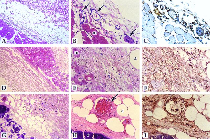
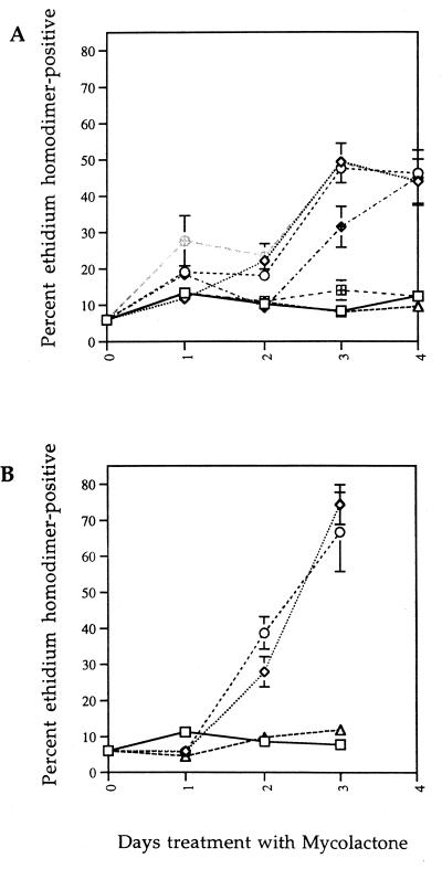
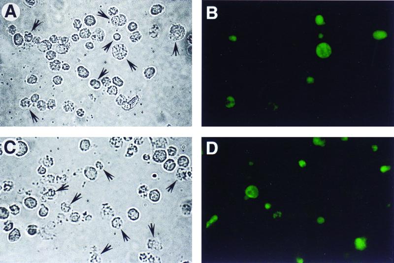
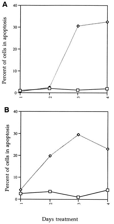
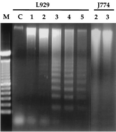
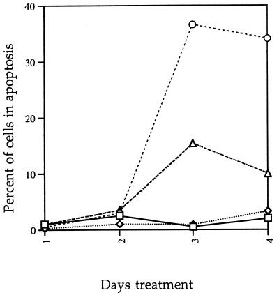
Mycobacterium ulcerans is the causative agent of Buruli ulcer, a tropical ulcerative skin disease. One of the most intriguing aspects of this disease is the presence of extensive tissue damage in the absence of an acute inflammatory response. We recently purified and characterized a macrolide toxin, mycolactone, from M. ulcerans. Injection of this molecule into guinea pig skin reproduced cell death and lack of acute inflammatory response similar to that seen following the injection of viable bacteria. We also showed that mycolactone causes a cytopathic effect on mouse fibroblast L929 cells that is characterized by cytoskeletal rearrangements and growth arrest within 48 h. However, these results could not account for the extensive cell death which occurs in Buruli ulcer. The results presented here demonstrate that L929 and J774 mouse macrophage cells die via apoptosis after 3 to 5 days of exposure to mycolactone. Treatment of cells with a pan-caspase inhibitor can inhibit mycolactone-induced apoptosis. We demonstrate that injection of mycolactone into guinea pig skin results in cell death via apoptosis and that the extent of apoptosis increases as the lesion progresses. These results may help to explain why tissue damage in Buruli ulcer is not accompanied by an acute inflammatory response.


Figures

FIG. 1
FIG. 1
Detection of apoptosis in M. ulcerans-infected guinea pig lesions and media controls. The TUNEL reaction was performed, detected by use of DAB, and counterstained with hematoxylin as described in Materials and Methods. A to C, negative control section injected with mycobacterial medium; D to F, M. ulcerans-injected section (8 days postinjection). A and D, ×83; B, C, E, and F, ×332. Panels A, B, D, and E are hematoxylin-eosin-stained sections; panels C and F are TUNEL sections. Arrows point to the blood vessels; muscle cells (m) are also indicated.
FIG. 2
FIG. 2
Detection of apoptosis in mycolactone-induced guinea pig lesions. The TUNEL reaction was performed, detected by use of DAB, and counterstained with hematoxylin-eosin as described in Materials and Methods. The mycobacterial-medium control is shown in panels A to C of Fig. 1. A to C, 2 days p.i.; D to F, 8 days p.i; G to I, 22 days p.i. Magnifications: A, D, and G, ×83; B, C, E, F, H, and I, ×332. Panels A, B, D, E, G, and H are hematoxylin-eosin-stained sections; panels C, F, and I are TUNEL sections. Arrows point to blood vessels; muscle (m), adipocytes (a), and calcification (c) are also indicated.
FIG. 3
FIG. 3
Mycolactone-mediated cell death of L929 and J774 cells. (A) Cytoxicity on L929 cells. L929 cells were treated with mycolactone at 30 pg/ml (hatched squares), 300 pg/ml (open triangles), 3 ng/ml (hatched diamonds), 30 ng/ml (open diamonds), 300 ng/ml (hatched circles), 3 μg/ml (open circles), or ethanol alone (open squares). (B) Cytoxicity on J774 cells. J774 cells were treated with mycolactone at 300 pg/ml (open triangles), 30 ng/ml (open diamonds), 3 μg/ml (open circles), or ethanol alone (open squares). Cell death was measured by ethidium homodimer uptake as described in Materials and Methods.
FIG. 4
FIG. 4
TUNEL reaction of L929 and J774 cells. (A and B) The TUNEL reaction was performed on L929 cells treated with mycolactone at 300 ng/ml for 3 days. (C and D) TUNEL reaction was performed on J774 cells treated with mycolactone at 300 ng/ml for 3 days. Panels A and C are phase-contrast micrographs, and panels B and D are fluorescence micrographs. Arrows in panels A and C identify the TUNEL-positive cells.
FIG. 5
FIG. 5
Quantitation and kinetic analysis of apoptotic L929 and J774 cells. The TUNEL reaction was performed on L929 (A) and J774 (B) cells and quantitated as described in Materials and Methods. Symbols: □, control (ethanol-treated) cells; ◊, mycolactone-treated cells.
FIG. 6
FIG. 6
Electrophoretic analysis of DNA from L929 and J774 cells exposed to mycolactone. Genomic DNA was extracted from cells treated with 300 ng of mycolactone per ml and electrophoresed on a 1.5% agarose gel. Lane M, molecular weight markers; lane C, control L929 cells (treated with ethanol and harvested at day 5). Numbers above the lanes indicate days of treatment with mycolactone.
FIG. 7
FIG. 7
B-D-FMK inhibits mycolactone-induced apoptosis. L929 cells were treated with ethanol only (□), 1 μM B-D-FMK only (◊), mycolactone only (○), or mycolactone plus B-D-FMK (▵). Cells were harvested at each day and processed for the TUNEL reaction.

References

    1. Aagaard-Tillery K M, Jelinek D F. Inhibition of human B lymphocyte cell cycle progression and differentiation by rapamycin. Cell Immunol. 1994;156:493–507. - PubMed
    1. Aagaard-Tillery K M, Jelinek D F. Differential activation of a calcium-dependent endonuclease in human B lymphocytes. Role in ionomycin-induced apoptosis. J Immunol. 1995;155:3297–3307. - PubMed
    1. Aoshiba K, Nagai A, Konno K. Erythromycin shortens neutrophil survival by accelerating apoptosis. Antimicrob Agents Chemother. 1995;39:872–877. - PMC - PubMed
    1. Bryskier A, Agouridas C, Gasc J-C. Classification of macrolide antibiotics. In: Bryskier A J, Butzler J-P, Neu H C, Tulkens P M, editors. Macrolides: chemistry, pharmacology and clinical uses. Paris, France: Arnette S.A.; 1993. pp. 5–66.
    1. Cohen G M, Sun X-M, Snowden R T, Dinsdale D, Skilleter D N. Key morphological features of apoptosis may occur in the absence of internucleosomal DNA fragmentation. Biochem J. 1992;286:331–334. - PMC - PubMed

LinkOut - more resources