. 2000 Jan 1;14(1):34-44.

Regulation of tumor angiogenesis by p53-induced degradation of hypoxia-inducible factor 1alpha

Affiliations

Regulation of tumor angiogenesis by p53-induced degradation of hypoxia-inducible factor 1alpha

R Ravi et al. Genes Dev. .

Abstract

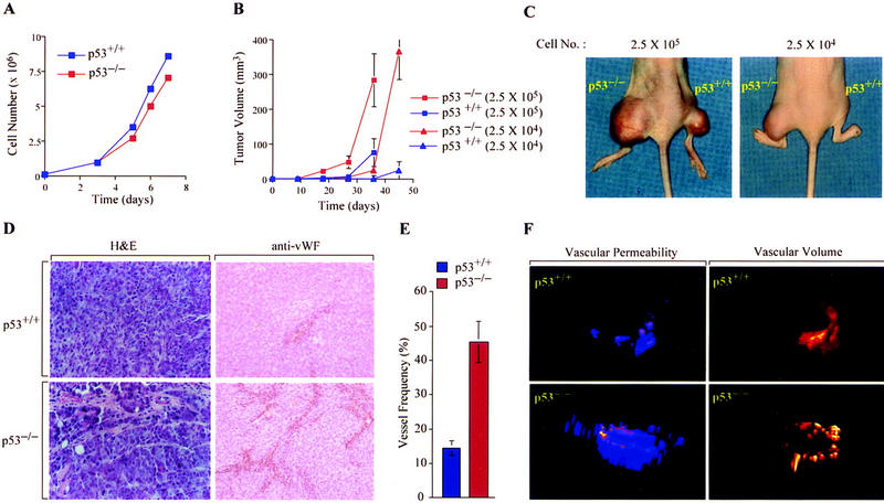
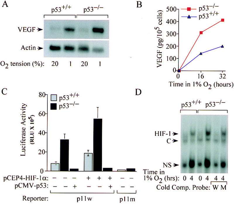
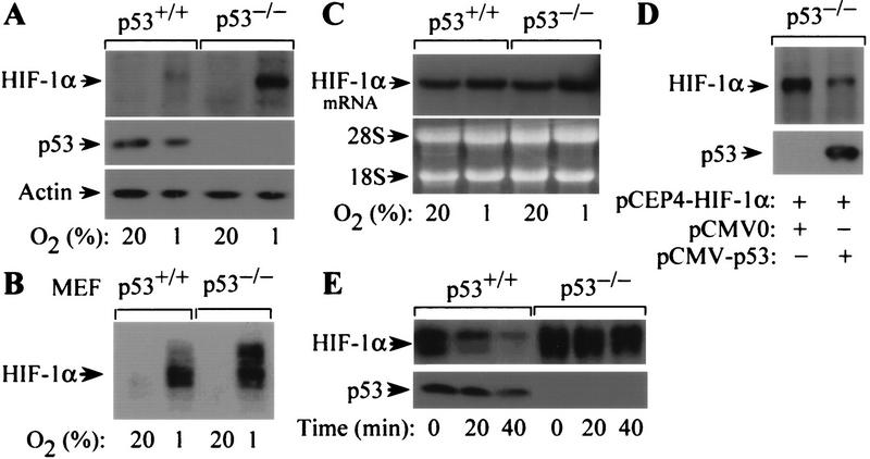
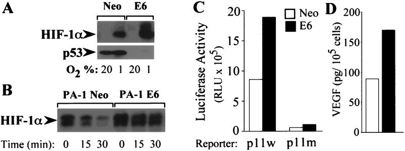
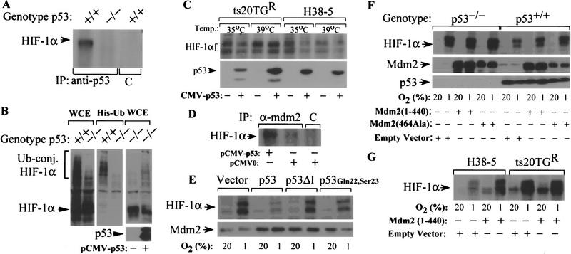
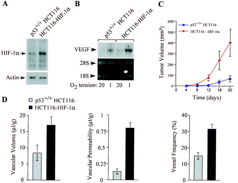
The switch to an angiogenic phenotype is a fundamental determinant of neoplastic growth and tumor progression. We demonstrate that homozygous deletion of the p53 tumor suppressor gene via homologous recombination in a human cancer cell line promotes the neovascularization and growth of tumor xenografts in nude mice. We find that p53 promotes Mdm2-mediated ubiquitination and proteasomal degradation of the HIF-1alpha subunit of hypoxia-inducible factor 1 (HIF-1), a heterodimeric transcription factor that regulates cellular energy metabolism and angiogenesis in response to oxygen deprivation. Loss of p53 in tumor cells enhances HIF-1alpha levels and augments HIF-1-dependent transcriptional activation of the vascular endothelial growth factor (VEGF) gene in response to hypoxia. Forced expression of HIF-1alpha in p53-expressing tumor cells increases hypoxia-induced VEGF expression and augments neovascularization and growth of tumor xenografts. These results indicate that amplification of normal HIF-1-dependent responses to hypoxia via loss of p53 function contributes to the angiogenic switch during tumorigenesis.


Figures

Figure 1
Figure 1
Effect of p53 genotype on tumor growth and angiogenesis. (A) Growth of p53+/+ (blue) and p53−/− (red) HCT116 cells cultured in DMEM supplemented with 10% fetal calf serum at 37°C and 95%air/5%CO2. (B, C) Growth of p53+/+ (blue) and p53−/− (red) HCT116 xenografts [2.5 × 104 (▴) or 2.5 × 105 (█) cells] injected subcutaneously into right (p53+/+) or left (p53−/−) hind legs of athymic BALB/c (nu/nu) mice. Values expressed represent mean ± s.e. of 12 xenografts of each cell type. (D) Histologic analysis of blood vessels in p53+/+ and p53−/− HCT116 xenograft tumors by staining with H&E or immunoperoxidase detection of endothelial cells using an anti-vWF antibody (×25). (E) Quantification of blood vessel density in p53+/+ (blue) and p53−/− (red) xenografts. The data represent the mean ± s.e. of the frequency of vessel hits among 300 random sampling points from each of three tumors of either genotype. (F) Representative NMR analysis of in vivo vascular volume (right) and permeability (left) of p53+/+ and p53−/− (bottom) HCT116 xenografts.
Figure 2
Figure 2
Effect of p53 genotype on hypoxia-induced VEGF expression and HIF-1 activity. (A) Northern blot analysis of VEGF mRNA expression in p53+/+ and p53−/− HCT116 cells incubated for 16 hr in either 20%or 1%O2. (B) ELISA of VEGF protein concentration in supernatant medium of p53+/+ (blue ▴) or p53−/− (red █) HCT116 cells incubated for 16–32 hr in 1% O2. (C) Hypoxia-induced and HIF-1-dependent activation of VEGF-reporter activity in p53+/+ (shaded bars) and p53−/− (solid bars) HCT116 cells. Wild-type (p11w) and mutant (p11m) copies of the hypoxia response element from the VEGF gene were inserted 5′ to a SV40 promoter–luciferase transcription unit. Cells were cotransfected with either VEGF–p11w or VEGF–p11m and CMVβgal, with or without pCEP4/HIF-1α or pCMV–p53, exposed to 1% O2 for 20 hr, and harvested for luciferase assays. The data represent the mean ± s.e. luciferase activity (normalized for β-gal activity) from three independent experiments. (D) Electrophoretic mobility shift assays of HIF-1 DNA-binding activity in nuclear extracts from p53+/+ and p53−/− HCT116 cells exposed to 20% (lanes 1 and 3) or 1% (lanes 2 and 4–6) O2. HIF-1 DNA binding was confirmed by competition assays using either unlabeled wild-type oligonucleotide (W) or a mutant oligonucleotide (M) containing the same 3-bp substitution as in p11m. Complexes containing HIF-1, constitutive (C), and nonspecific (NS) DNA-binding activities (Semenza and Wang 1992) are indicated.
Figure 3
Figure 3
Effect of p53 on oxygen-regulated expression and stability of HIF-1α. (A) Immunoblot analysis of HIF-1α expression in p53+/+ and p53−/− HCT116 cells cultured for 8 hr in 20%or 1%O2. The blot was analyzed sequentially with monoclonal antibodies against HIF-1α (H1α67), p53 (DO-1), and β-actin. (B) Immunoblot analysis of HIF-1α expression in p53+/+ and p53−/− MEFs cultured for 8 hr in 20% or 1% O2. (C) Northern blot analysis of HIF-1α mRNA expression in p53+/+ and p53−/− HCT116 cells cultured as in A. (D). Immunoblot analysis of HIF-1α protein in p53−/− HCT116 cells cultured in 1% O2 for 8 hr following cotransfection with pCEP4–HIF-1α and either pCMV–p53 or empty vector. The blot was analyzed sequentially with anti-HIF-1α and anti-p53 monoclonal antibodies. (E) Half-life of HIF-1α protein in p53+/+ and p53−/− cells exposed to 100 μm cobalt chloride following addition of 100 μm cycloheximide. Lysates of cells harvested at the indicated time intervals were subject to immunoblot analysis of HIF-1α and p53 expression.
Figure 4
Figure 4
HPV E6 increases expression of HIF-1α and VEGF in response to hypoxia. (A) Immunoblot analysis of HIF-1α expression in PA-1 Neo or PA-1 E6 cells cultured for 8 hr in 20% or 1% O2. (B) Half-life of HIF-1α protein in PA-1 Neo or PA-1 E6 cells exposed to 100 μm cobalt chloride following addition of 100 μm cycloheximide. Lysates of cells harvested at the indicated time intervals were subject to immunoblot analysis of HIF-1α expression. (C) Hypoxia-induced and HIF-1-dependent activation of VEGF-reporter activity in PA-1 Neo (open bars) and PA-1 E6 (solid bars) cells. Cells were cotransfected with either VEGF–p11w or VEGF–p11m and CMVβgal, exposed to 1% O2 for 20 hr, and harvested for luciferase assays. The data represent the mean luciferase activity (normalized for β-gal activity) from three independent experiments. (D) ELISA of VEGF protein concentration in supernatant medium of PA-1 Neo (open bar) or PA-1 E6 (solid bar) cells incubated for 16 hr in 1% O2.
Figure 5
Figure 5
Effect of p53 expression on ubiquitin-mediated degradation of HIF-1α. (A) Interaction of p53 with HIF-1α. Lysates of p53+/+ or p53−/− HCT116 cells exposed to 1%O2 for 8 hr were immunoprecipitated with either anti-p53 antibody or isotype control antibody (C) and the resultant immune complexes were subjected to immunoblot analysis with anti-HIF-1α monoclonal antibody. (B) Differential ubiquitination of HIF-1α in hypoxic p53+/+ and p53−/− HCT116 cells. Cells were cotransfected with pCMVβgal and pCEP4/HIF-1α with either MT107/His6-Ub or empty vector (MT107), and cultured in 1%O2 for 4 hr in the presence of 50 μm MG132. Aliquots of whole-cell extract (WCE) or His-tagged proteins purified from whole-cell lysates (His-Ub) were subjected to immunoblot analysis with anti-HIF-1β antibody. (C) Effect of p53 expression on HIF-1α protein levels in hypoxic ts20TGR and H38-5 cells. Cells transfected with pCMV–p53 or pCMVβgal were maintained at either 35°C or 39°C for 8 hr and exposed to 1%O2 for an additional 8 hr at their respective temperatures. Whole-cell lysates were subjected to immunoblot analysis with anti-HIF-1α or anti-p53 antibodies. (D) Effect of p53 on complex formation between HIF-1α and Mdm2. Lysates of p53−/− HCT116 cells transfected with either pCMV–p53 or empty vector and transferred to 1%O2 for 6 hr were immunoprecipitated with anti-Mdm2 or isotype control antibody, and the resulting immune complexes were subjected to immunoblot assays using an antibody against HIF-1α. (E) Effect of wild-type p53, p53ΔI, or p53Gln22,Ser23 on expression of HIF-1α in response to hypoxia. p53−/− HCT116 cells transfected with pCMVβgal and either pCMV–p53, pCB6 + p53ΔI, pCMV–p53Gln22,Ser23, or empty vector were exposed to 1%O2 for 8 hr. Whole-cell lysates were subjected to immunoblot analysis with anti-HIF-1α or anti-Mdm2 antibodies. (F) Effect of dominant–negative (RING finger) mutants of Mdm2 on hypoxia-induced expression of HIF-1α. p53+/+ and p53−/− HCT116 cells transfected with vectors encoding human Mdm2 (1–440) (pCHDM1–440), Mdm2 (464Ala) (pCHDM464Ala), or pCMVβgal were exposed to 1%O2 for 8 hr. Whole-cell lysates were subjected to immunoblot analysis with anti-HIF-1α, anti-p53, or anti-Mdm2 antibodies. (G) Effect of dominant–negative (RING finger deletion mutant) Mdm2 on p53-mediated inhibition of HIF-1α expression in ts20TGR and H38-5 cells. Cells cotransfected with pCMV–p53 and either pCHDM1–440 or empty vector were maintained at 39°C for 12 hr and then exposed to 20%or 1%O2 for an additional 8 hr at 39°C. Whole-cell lysates were subjected to anti-HIF-1α immunoblot analysis.
Figure 6
Figure 6
Increased tumor angiogenesis and growth in p53+/+ cells with forced overexpression of HIF-1α. (A) Immunoblot analyses of HIF-1α protein levels in p53+/+ HCT116 cells and p53+/+ HCT116 cells stably transfected with a HIF-1α expression vector (HCT116–HIF-1α following exposure to 1%O2 for 8 hr. (B) Northern blot analysis of VEGF mRNA levels in p53+/+ HCT116 and HCT116–HIF-1α cells cultured for 16 hr in 20%or 1%O2. (C) Growth of p53+/+ HCT116 (blue █) and HCT116–HIF-1α (red ▴) cells (2.5 × 106) injected subcutaneously into the flanks of athymic BALB/c nude mice. Values expressed represent mean ± s.e. of 12 xenografts of each cell type. (D) Quantification of vascular volume, permeability, and blood vessel density in p53+/+ HCT116 (shaded bars) and HCT116–HIF-1α (solid bars) xenograft tumors. In vivo vascular volume and permeability of the tumors were determined by NMR analyses, and blood vessel frequency in stained sections of excised tumors was analyzed as described in Fig. 1.

References

    1. An WG, Kanekal M, Simon MC, Maltepe E, Blagosklonny MV, Neckers LM. Stabilization of wild-type p53 by hypoxia-inducible factor 1α. Nature. 1998;392:405–408. - PubMed
    1. Barak Y, Juven T, Haffner R, Oren M. Mdm-2 expression is induced by wild type p53 activity. EMBO J. 1993;12:461–468. - PMC - PubMed
    1. Bergers G, Javaherian K, Lo KN, Folkman J, Hanahan D. Effects of angiogenesis inhibitors on multistage carcinogenesis in mice. Science. 1999;284:808–812. - PubMed
    1. Blagosklonny MV, An WG, Romanova LY, Trepel J, Fojo T, Neckers L. p53 inhibits hypoxia-inducible factor-stimulated transcription. J Biol Chem. 1998;273:11995–11998. - PubMed
    1. Boehm T, Folkman J, Browder T, O'Reilly MS. Antiangiogenic therapy of experimental cancer does not induce acquired drug resistance. Nature. 1997;390:404–407. - PubMed

Publication types

MeSH terms