. 2000 Jan 1;14(1):108-17.

HAESA, an Arabidopsis leucine-rich repeat receptor kinase, controls floral organ abscission

Affiliations

HAESA, an Arabidopsis leucine-rich repeat receptor kinase, controls floral organ abscission

T L Jinn et al. Genes Dev. .

Abstract

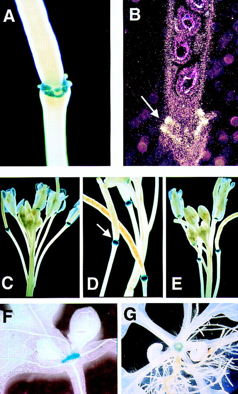
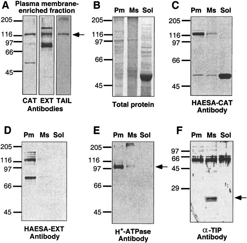
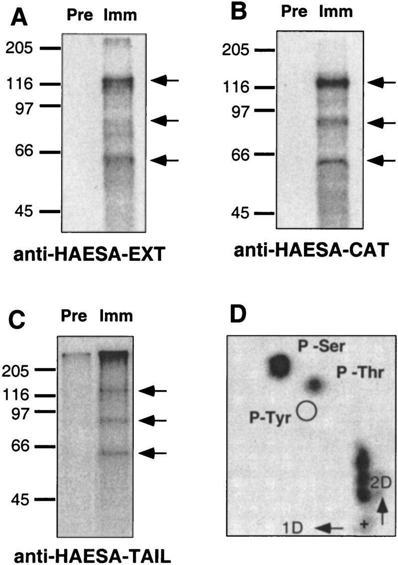
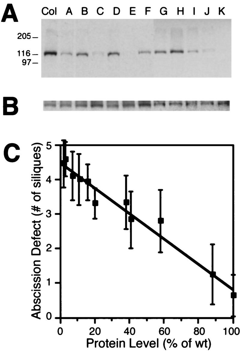
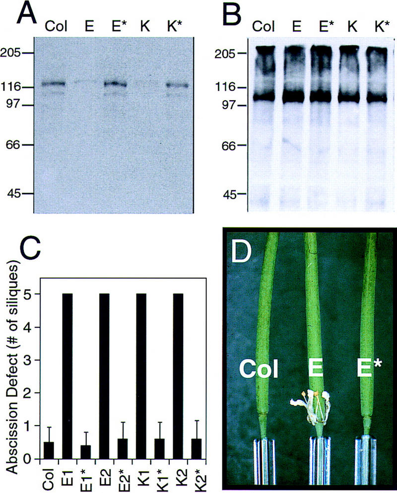
Abcission, the natural shedding of leaves, flowers and fruits, is a fundamental component of plant development. Abscission is a highly regulated process that occurs at distinct zones of cells that undergo enlargement and subsequent separation. Although some components of abscission, including accumulation of the hormone ethylene and cell wall-degrading enzymes, have been described, the regulatory pathways remain largely unknown. In this paper we describe a critical component required for floral organ abscission in Arabidopsis thaliana, the receptor-like protein kinase HAESA. Histochemical analysis of transgenic plants harboring a HAESA promoter:: beta-glucuronidase reporter gene and in situ RNA hybridization experiments show HAESA expression in the abscission zones where the sepals, petals, and stamens attach to the receptacle, at the base of pedicels, and at the base of petioles where leaves attach to the stem. Immunodetection, immunoprecipitation, and protein kinase activity assays reveal HAESA is a plasma membrane serine/threonine protein kinase. The reduction of function of HAESA in transgenic plants harboring an antisense construct results in delayed abscission of floral organs, and the severity of the phenotype is directly correlated with the level of HAESA protein. These results demonstrate that HAESA functions in developmentally regulated floral organ abscission.


Figures

Figure 1
Figure 1
HAESA expression pattern. (A) Transgenic plants harboring the HAESA promoter fused to the GUS reporter gene were histochemically stained for GUS activity using the chromogenic substrate 5-bromo-4-chloro-3 indolyl glucuronide. GUS activity (indicated by blue color) at the base of the flowers shows that HAESA expression is restricted to the abscission zones, in which the sepals, petals, and stamens have detached. (B) In situ RNA hybridization confirms the results of the HAESA::GUS expression in reproductive tissues. Longitudinal sections of a mature silique were hybridized with a HAESA antisense probe. Silver grains deposited after development of the photographic film (indicated by the small white-colored dots) correspond to HAESA mRNA and can be seen at the base of the silique (arrow). No detectable signal was observed in similar sections hybridized with a HAESA sense probe. (C) Developmental stage-specific HAESA expression, in which the first flower competent for pollination shows GUS activity at the base of the flower. (D) Pollination-independent HAESA::GUS expression. An emasculated flower in which the anthers were removed prior to dehiscence still shows GUS activity (arrow, unpollinated silique without seeds). (E) HAESA::GUS expression in the F1 hybrid derived from etr1-1 and the HAESA::GUS transgenic, showing that HAESA expression is independent of ethylene signal transduction. HAESA::GUS expression was also observed in vegetative tissues. (F) A side view of a seedling at the four-leaf stage; (G) a view from the top of a seedling at the six- to eight-leaf stage. GUS activity is restricted to a donut-shaped region at the base of the petioles where the leaves attach to the stem.
Figure 2
Figure 2
Subcellular immunolocalization of HAESA. Three different polyclonal antibodies were raised against portions of recombinant HAESA protein (CAT, EXT, and TAIL) and affinity purified for Western blot analyses. Protein extracts from mature rosettes were fractionated using a PEG/dextran two-phase partition system to obtain a plasma membrane-enriched (Pm), a total microsomal membrane (Ms) and a soluble protein (Sol) fraction. (A) A comparison of Western blot analyses using the three different antibodies against plasma membrane-enriched fractions (the arrow indicates the ∼120-kD protein recognized by all three HAESA antibodies); (B) total protein profiles assessed by Coommassie blue staining; and Western blot analyses of Pm, Ms, and Sol fractions using CAT (C) and EXT (D) antibodies. To illustrate the purity of the two-phase partitioned fractions, Western blot analyses with various fractions were performed with antibodies to the plasma membrane-specific H+–ATPase (E; the arrow indicates the ∼100-kD protein) and the tonoplast-specific α-TIP (F; the arrow indicates the ∼27-kD protein). Sizes of the molecular mass markers (kD) are indicated at left.
Figure 3
Figure 3
Immunoprecipitation and autophosphorylation of HAESA. Solubilized proteins from plasma membrane-enriched fractions obtained by two-phase partitioning were immunoprecipitated with affinity-purified antibodies from preimmune (Pre) or immune (Imm) sera raised against three different portions of the HAESA protein (A, EXT; B, CAT; and C, TAIL). Immunocomplexes were autophosphorylated in the presence of [γ-32P]ATP, resolved by SDS-PAGE, and exposed to autoradiographic film. Sizes of the molecular mass markers are indicated at left; the HAESA 120-kD protein and ∼85- and ∼65-kD unknown polypeptides are indicated with arrows. Phosphoamino acid content was determined by isolation of the 120-kD phosphoprotein, acid-hydrolysis, and separation by two-dimensional thin-layer electrophoresis (D). The sample origin is indicated by +, and positions of phosphoamino acid standards visualized by staining with ninhydrin are circled. Incompletely hydrolyzed peptides are evident above the origin, and radioactive phosphoserine (P-Ser) and phosphothreonine (P-Thr) were detected, but phosphotyrosine (P-Tyr) was not.
Figure 4
Figure 4
Antisense suppression of HAESA and floral organ abscission phenotype. (A) Transgenic lines containing single copies of the strong, constitutive 35S cauliflower mosaic virus (CaMV) promoter driving an antisense version of HAESA cDNA were generated, and the levels of HAESA protein determined by Western blot analysis using the TAIL antibody. The parental wild-type (Col) is compared with 11 independent lines (indicated above each lane). Sizes of molecular mass markers (kD) are indicated at left. (B) An antibody raised against the plasma membrane-specific H+–ATPase (DeWitt et al. 1996) was used as a protein loading control. Densitometric quantitation indicates that the transgenic lines have suppressed levels of HAESA protein ranging from <10% of wild type (lines C, E, and K) to 88% of wild type (line H). (C) Floral organ abscission in the five most mature siliques were scored. Those that had failed to detach their floral organs were scored as defective. The number of siliques that failed to abscise floral organs (±s.d.) was plotted against the level of HAESA protein in wild type (Col), and the various antisense-suppressed transgenic lines determined by densitometry. A direct, inverse correlation between the amount of HAESA protein and the number of siliques in which floral organs failed to abscise was observed (r2 = 0.964). Col n = 150 flowers; antisense transgenic lines n = 45 flowers each.
Figure 5
Figure 5
Loss of the transgene leads to loss of the floral organ abscission phenotype. To confirm that the floral organ abscission defect observed in antisense-suppressed transgenic lines is due to the presence of the transgene, HAESA protein levels in wild-type (Col) and siblings that retain the transgene (E and K) or have lost the transgene (E* and K*) were determined by Western blot analysis using the TAIL antibody (A). (B) A duplicate Western blot probed with the plasma membrane H+–ATPase antibody ensures equal loading. Sizes of molecular mass markers (kD) are indicated at left. (C) Floral organ abscission (no. of siliques ±s.d.) of these segregating plants. Those plants that no longer carry the transgene (indicated by * ) have normal floral organ abscission. Col n = 100 flowers; lines derived from primary transformants E and K n = 60 flowers each. (D) A representative example of siliques from wild-type Col (left), antisense trangenic line E (middle; for clarity, the front sepal and petal are removed), and a sibling that had lost the transgene (E*; right).

References

    1. Altmann T. Recent advances in brassinosteroid molecular genetics. Curr Opin Plant Biol. 1998;1:378–383. - PubMed
    1. An G, Ebert PR, Mitra A, Ha SB. Binary vectors. Plant Mol Biol Manual. 1988;A3:1–19.
    1. Bechtold N, Bouchez D. In planta Agrobacterium-mediated transformation of adult Arabidopsis thaliana plants by vacuum infiltration. In: Potrykus I, Spangenberg G, editors. Gene transfer to plants. New York, NY: Springer Verlag; 1994. p. 361. - PubMed
    1. Becraft PW. Receptor kinases in plant development. Trends Plant Sci. 1998;3:384–388.
    1. Bleecker AB, Patterson SE. Last exit: Senescence, abscission, and meristem arrest in Arabidopsis. Plant Cell. 1997;9:1169–1179. - PMC - PubMed

Publication types

MeSH terms

Substances