

Ion channels and vascular tone
- PMID: 10642294
- PMCID: PMC1382026
- DOI: 10.1161/01.hyp.35.1.173
Ion channels and vascular tone
Abstract
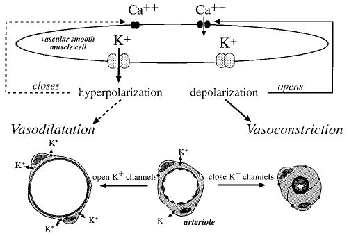
Ion channels in the plasma membrane of vascular muscle cells that form the walls of resistance arteries and arterioles play a central role in the regulation of vascular tone. Current evidence indicates that vascular smooth muscle cells express at least 4 different types of K(+) channels, 1 to 2 types of voltage-gated Ca(2+) channels, >/=2 types of Cl(-) channels, store-operated Ca(+) (SOC) channels, and stretch-activated cation (SAC) channels in their plasma membranes, all of which may be involved in the regulation of vascular tone. Calcium influx through voltage-gated Ca(2+), SOC, and SAC channels provides a major source of activator Ca(2+) used by resistance arteries and arterioles. In addition, K(+) and Cl(-) channels and the Ca(2+) channels mentioned previously all are involved in the determination of the membrane potential of these cells. Membrane potential is a key variable that not only regulates Ca(+2) influx through voltage-gated Ca(2+) channels, but also influences release of Ca(2+) from internal stores and Ca(2+)- sensitivity of the contractile apparatus. By controlling Ca(2+) delivery and membrane potential, ion channels are involved in all aspects of the generation and regulation of vascular tone.
Figures


References
-
- Nelson MT, Patlak JB, Worley JF, Standen NB. Calcium channels, potassium channels, and voltage dependence of arterial smooth muscle tone. Am J Physiol Cell Physiol. 1990;259:C3–C18. - PubMed
-
- Hughes AD. Calcium channels in vascular smooth muscle cells. J Vasc Res. 1995;32:353–370. - PubMed
-
- Yamagishi T, Yanagisawa T, Taira N. K+ channel openers, cromakalim and Ki4032, inhibit agonist-induced Ca2+ release in canine coronary artery. Naunyn Schmiedebergs Arch Pharmacol. 1992;346:691–700. - PubMed
-
- Kukuljan M, Rojas E, Catt KJ, Stojilkovic SS. Membrane potential regulates inositol 1,4,5-trisphosphate-controlled cytoplasmic Ca2+ oscillations in pituitary gonadotrophs. J Biol Chem. 1994;269:4860–4865. - PubMed
Publication types
MeSH terms
Substances
Grants and funding
LinkOut - more resources
Full Text Sources
Miscellaneous
