. 2000 Jan;105(2):233-9.
doi: 10.1172/JCI8691.

Deficiency of dolichol-phosphate-mannose synthase-1 causes congenital disorder of glycosylation type Ie

Affiliations

Deficiency of dolichol-phosphate-mannose synthase-1 causes congenital disorder of glycosylation type Ie

T Imbach et al. J Clin Invest. 2000 Jan.

Abstract

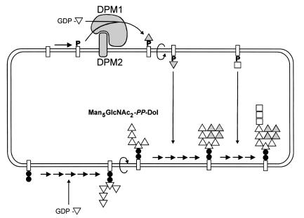
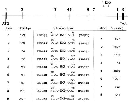
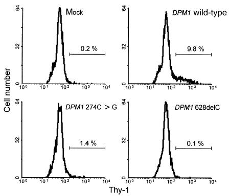
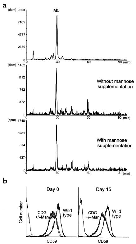
Congenital disorders of glycosylation (CDG), formerly known as carbohydrate-deficient glycoprotein syndromes, lead to diseases with variable clinical pictures. We report the delineation of a novel type of CDG identified in 2 children presenting with severe developmental delay, seizures, and dysmorphic features. We detected hypoglycosylation on serum transferrin and cerebrospinal fluid beta-trace protein. Lipid-linked oligosaccharides in the endoplasmic reticulum of patient fibroblasts showed an accumulation of the dolichyl pyrophosphate Man(5)GlcNAc(2) structure, compatible with the reduced dolichol-phosphate-mannose synthase (DolP-Man synthase) activity detected in these patients. Accordingly, 2 mutant alleles of the DolP-Man synthase DPM1 gene, 1 with a 274C>G transversion, the other with a 628delC deletion, were detected in both siblings. Complementation analysis using DPM1-null murine Thy1-deficient cells confirmed the detrimental effect of both mutations on the enzymatic activity. Furthermore, mannose supplementation failed to improve the glycosylation status of DPM1-deficient fibroblast cells, thus precluding a possible therapeutic application of mannose in the patients. Because DPM1 deficiency, like other subtypes of CDG-I, impairs the assembly of N-glycans, this novel glycosylation defect was named CDG-Ie.


Figures

Figure 1
Figure 1
Assembly of the oligomannose core in the ER. Mannose (triangles) is first incorporated into dolichol-bound oligosaccharides (rectangles) from the GDP-Man donor substrate. After flipping into the luminal side, Man5GlcNAc2-PP-Dol is further decorated with 4 mannoses provided by DolP-Man. The formation of DolP-Man from GDP-Man is catalyzed by the DolP-Man synthase enzyme, which is constituted of a catalytic subunit (Dpm1) and a membrane-anchored subunit (Dpm2). Once fully mannosylated, the core receives 3 glucose residues (squares) before being transferred by the oligosaccharyltransferase complex onto nascent glycoproteins. Filled circles, GlcNAc.
Figure 2
Figure 2
Isoelectric focusing and Western blotting. (a) Serum transferrin was separated by isoelectric focusing. The anode is at the top. The number of negative charges is indicated at the right. (b) β-trace protein was analyzed by SDS-PAGE followed by Western blotting. Molecular weights (in kDa) are given at the right. (Sib1 and sib2 are described in Table 1.)
Figure 3
Figure 3
Lipid-linked oligosaccharide profile in a CDG-Ie patient. [3H]oligosaccharides were separated by HPLC after labeling CDG fibroblasts with [3H]mannose for 1 hour. The top profile shows the retention times of yeast-derived oligosaccharide standards from Man1GlcNAc2 (M1) to Glc3Man9GlcNAc2 (G3M9). The bottom profile shows the Man5GlcNAc2 peak accumulating in the fibroblasts from a CDG-Ie patient.
Figure 4
Figure 4
Two mutant alleles of the DPM1 gene in CDG-Ie patients. The positions of the 274C>G and 628delC mutations are marked in black on the human DPM1 cDNA sequence. The early termination of DPM1 translation caused by the 628delC-mediated frameshift is shown as a vertical line at position 640 of the cDNA.
Figure 5
Figure 5
Genomic organization of the human DPM1 gene. Exons are numbered and represented as filled rectangles. Introns are indicated as solid lines. The positions of initiation and termination of translation are symbolized by ATG and TAA, respectively. The sizes of the exons and introns are shown proportionally. The bottom panel shows the sizes and boundaries of the DPM1 exons and introns.
Figure 6
Figure 6
Complementation of Thy1-E cells. BW5147 Thy1-null cells, transfected with DPM1 expression vectors, were analyzed for Thy1 staining by flow cytometry. Each panel shows the fluorescence histogram obtained for cells transfected with a mock vector, the human DPM1wild-type cDNA, the human DPM1 274C>G cDNA, and the DPM1 628delC cDNA. The region marked with the horizontal bar denotes the percentage of Thy1-positive cells.
Figure 7
Figure 7
Mannose supplementation in CDG fibroblasts. Lipid-linked oligosaccharide profiles (a) and CD59 levels (b) were analyzed in CDG cells cultured for 15 days in the presence of 5 mM mannose. The top panel shows the HPLC elution profile of the Man5GlcNAc2 standard (M5) derived from the Saccharomyces cerevisiae alg3 yeast mutant cells.

Comment in

References

    1. Jaeken J, Carchon H, Stibler H. The carbohydrate-deficient glycoprotein syndromes: pre-Golgi and Golgi disorders? Glycobiology. 1993;3:423–428. - PubMed
    1. Krasnewich D, Gahl WA. Carbohydrate-deficient glycoprotein syndrome. Adv Pediatr. 1997;44:109–140. - PubMed
    1. Varki A. Biological roles of oligosaccharides: all of the theories are correct. Glycobiology. 1993;3:97–130. - PMC - PubMed
    1. Matthijs G, et al. Mutations in PMM2, a phosphomannomutase gene on chromosome 16p13, in carbohydrate-deficient glycoprotein type I syndrome (Jaeken syndrome) Nat Genet. 1997;16:88–92. - PubMed
    1. Niehues R, et al. Carbohydrate-deficient glycoprotein syndrome type Ib. Phosphomannose isomerase deficiency and mannose therapy. J Clin Invest. 1998;101:1414–1420. - PMC - PubMed

Publication types

MeSH terms