

Immunohistochemical analysis of the distribution of measles related antigen in the intestinal mucosa in inflammatory bowel disease
- PMID: 10644308
- PMCID: PMC1727809
- DOI: 10.1136/gut.46.2.163
Immunohistochemical analysis of the distribution of measles related antigen in the intestinal mucosa in inflammatory bowel disease
Abstract
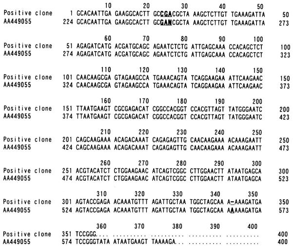
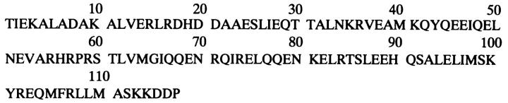
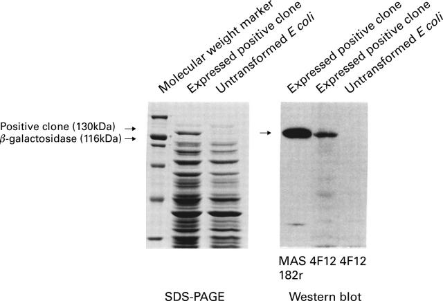
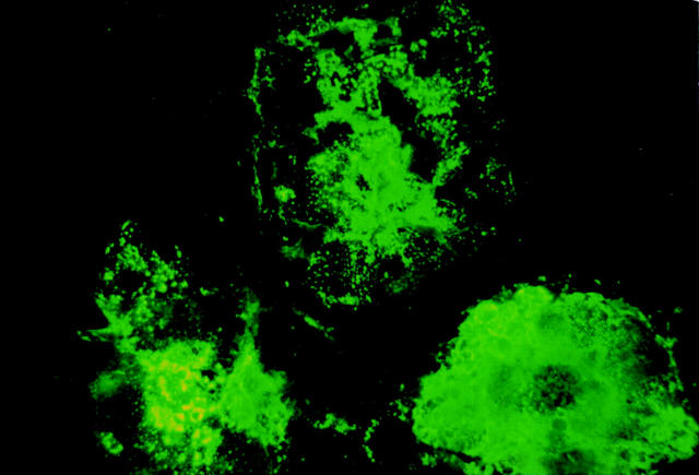
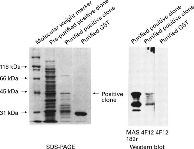
Background: Measles virus is implicated in the aetiology of Crohn's disease. This measles hypothesis is mainly supported by immunohistochemical findings that the measles related antigen is present in the intestine of patients with Crohn's disease. Recently we isolated this antigen from the intestine of a patient with Crohn's disease using a molecular cloning technique and produced the monoclonal antibody against it (designated 4F12).
Aim: To discover whether the measles related antigen is uniquely present in Crohn's disease.
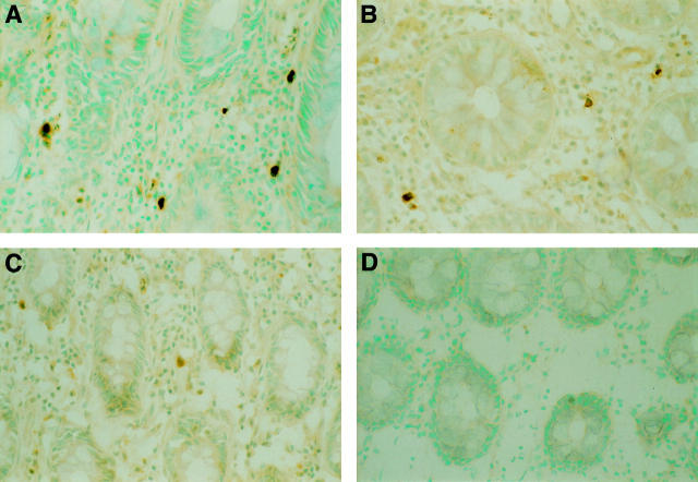
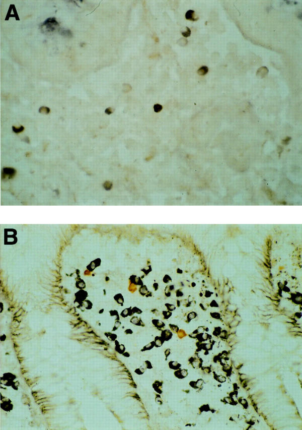
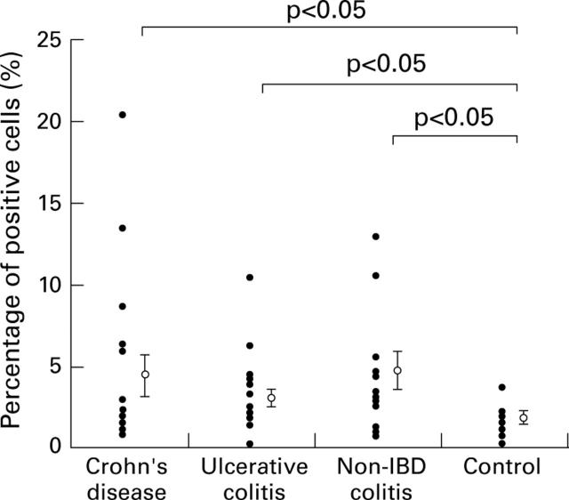
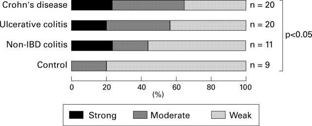
Subjects/methods: Colonic mucosa samples from 20 patients with Crohn's disease, 20 with ulcerative colitis, 11 with non-inflammatory bowel disease (IBD) colitis, and nine controls were immunohistochemically stained with the anti-measles monoclonal antibody 4F12. The numbers of positive cells, the ratio of positive cells to nucleated cells, and the staining intensity of the positive cells were compared. Furthermore, the distribution of the measles antigen in other human organs was examined.
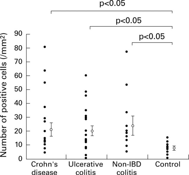
Results: Both the number of positive cells and the ratio of positive cells to nucleated cells were significantly increased in Crohn's disease, ulcerative colitis, and non-IBD colitis compared with controls (p<0.05) but were similar among the three disease groups. The staining intensity of the positive cells was also similar among the three disease groups. Small numbers of positive cells were observed in the oesophagus, stomach, duodenum, jejunum, and lung.
Conclusions: The presence of the measles related antigen in the colonic mucosa was not unique to Crohn's disease. These results, together with the observation that such a measles related antigen was derived from host protein, do not support the hypothesis that measles virus causes Crohn's disease.
Figures
Comment in
-
Immunohistochemical analysis of measles related antigen in IBD.Gut. 2001 Jan;48(1):136-7. doi: 10.1136/gut.48.1.136. Gut. 2001. PMID: 11200076 Free PMC article. No abstract available.
Similar articles
-
Detection of immunoreactive antigen, with a monoclonal antibody to measles virus, in tissue from a patient with Crohn's disease.J Gastroenterol. 1995 Feb;30(1):28-33. doi: 10.1007/BF01211371. J Gastroenterol. 1995. PMID: 7719411
-
Immunoglobulin G (IgG), IgG1, and IgG2 determinations from endoscopic biopsy specimens in control, Crohn's disease, and ulcerative colitis subjects.Gut. 1992 Apr;33(4):507-12. doi: 10.1136/gut.33.4.507. Gut. 1992. PMID: 1582596 Free PMC article.
-
Detection and comparative analysis of persistent measles virus infection in Crohn's disease by immunogold electron microscopy.J Clin Pathol. 1997 Apr;50(4):299-304. doi: 10.1136/jcp.50.4.299. J Clin Pathol. 1997. PMID: 9215145 Free PMC article.
-
[Distribution of cells containing different IgG and IgA subclasses in the colonic mucosa in childhood ulcerative colitis and Crohn disease].Orv Hetil. 1991 Sep 15;132(37):2027-31. Orv Hetil. 1991. PMID: 1923475 Review. Hungarian.
-
[Characteristics of HLA II antigen expression in the rectal mucosa of children suffering form ulcerative colitis and Crohn disease].Orv Hetil. 1995 Oct 15;136(42):2279-82. Orv Hetil. 1995. PMID: 7478471 Review. Hungarian.
Cited by
-
MMR: where are we now?Arch Dis Child. 2007 Dec;92(12):1055-7. doi: 10.1136/adc.2006.103531. Epub 2007 Jul 11. Arch Dis Child. 2007. PMID: 17626143 Free PMC article. Review.
-
Clinical aspects and pathophysiology of inflammatory bowel disease.Clin Microbiol Rev. 2002 Jan;15(1):79-94. doi: 10.1128/CMR.15.1.79-94.2002. Clin Microbiol Rev. 2002. PMID: 11781268 Free PMC article. Review.
-
No evidence of persistent mumps virus infection in inflammatory bowel disease.Gut. 2001 May;48(5):637-41. doi: 10.1136/gut.48.5.637. Gut. 2001. PMID: 11302960 Free PMC article.
-
Molecular mimicry, inflammatory bowel disease, and the vaccine safety debate.BMC Med. 2014 Sep 18;12:166. doi: 10.1186/s12916-014-0166-6. BMC Med. 2014. PMID: 25238056 Free PMC article.
-
Bioinformatic and immunological analysis reveals lack of support for measles virus related mimicry in Crohn's disease.BMC Med. 2014 Aug 28;12:139. doi: 10.1186/s12916-014-0139-9. BMC Med. 2014. PMID: 25168804 Free PMC article.
References
Publication types
MeSH terms
Substances
LinkOut - more resources
Full Text Sources
Medical