. 2000 Feb;46(2):182-90.
doi: 10.1136/gut.46.2.182.

Presence of sorbin in human digestive tract and endocrine digestive tumours

Affiliations

Presence of sorbin in human digestive tract and endocrine digestive tumours

F Abou El Fadil-Nicol et al. Gut. 2000 Feb.

Abstract

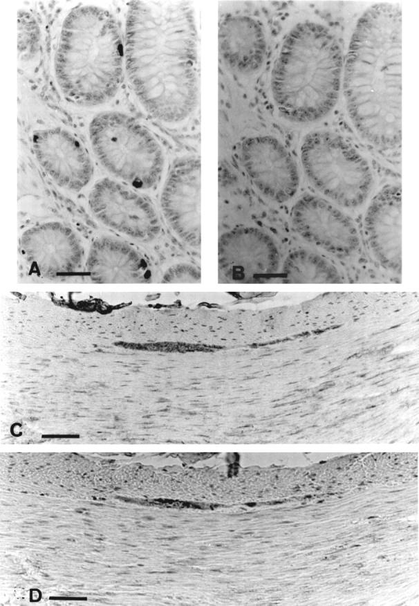
Background: Sorbin, a 153 amino acid peptide isolated from porcine intestine, was localised by immunohistochemistry in endocrine cells of the intestinal mucosa and pancreas and in the enteric nervous system in the pig.

Aims: To identify sorbin cells in normal human digestive tissues and to explore the expression of sorbin in 37 digestive endocrine tumours: 14 intestinal carcinoid tumours and 23 endocrine pancreatic tumours including six insulinomas.

Methods: Two polyclonal antibodies against the C-terminal and the N-terminal sequences of porcine sorbin raised in rabbit were used to evaluate sorbin expression by immunohistochemistry.

Results: In the human digestive tract, sorbin, characterised by both C-terminal and N-terminal immunoreactivity, was found in enterochromaffin cells of the gastric and intestinal epithelium from the pyloric junction to the descending colon. C-Terminal sorbin immunoreactivity alone was found in plexii from the enteric nervous system and in some insulin-containing cells of normal pancreas. C-Terminal and N-terminal antibodies disclosed sorbin in five of 14 intestinal carcinoid tumours; C-terminal antibody alone disclosed a C-terminal sorbin peptide in two of six insulinomas and three of 17 endocrine pancreatic tumours. The presence of sorbin was not associated with a specific clinical syndrome.

Conclusions: Sorbin is present in the digestive tract in several forms. It is expressed in some intestinal and pancreatic endocrine tumours.


Figures

Figure 1
Figure 1
Amino acid sequence of sorbin showing positions of synthetic peptides used for raising antibodies.
Figure 2
Figure 2
Localisation of sorbin immunoreactivity in the human duodenum. (A), (B), (C), and (D) are homologous fields of four adjacent sections. (A) Cells immunoreactive with the N-terminal sorbin antiserum. (B) Control, the immunoreactivity is partially blocked by adsorption on the homologous peptide, N8-18 peptide. (C) Cells immunoreactive with the C-terminal sorbin antiserum. (D) Control, the immunoreactivity is completely blocked by adsorption on the homologous peptide, Y-C17-NH2 peptide. Sorbin positive cells are located in the crypts of Lieberkühn. C-Terminal immunoreactive cells appear less numerous because of better specificity and the use of a higher dilution (1:500) of the C-terminal antiserum. (E) Sorbin-containing cells in Brunner's glands. Bouin's fixation. Visualisation with StreptABComplex/HRP. Haematoxylin counterstain. Scale bar = 50 µm.
Figure 3
Figure 3
Sorbin localisation in human ascending colon. (A) Sorbin-containing cells disclosed by the N-terminal antiserum in the colonic glands. (B) Control. On the adjacent section, the reaction was completely blocked after adsorption of the antiserum by the homologous peptide. (C) Immunoreactivity to C-terminal antiserum of a colonic myenteric plexus. (D) Colocalisation with chromogranin in the same plexus (adjacent section). Bouin's fixation. Visualisation with StreptABComplex/HRP. Haematoxylin counterstain. Scale bar = 50 µm.
Figure 4
Figure 4
Colocalisation of sorbin (A) and serotonin (B) in enterochromaffin cells of the human duodenum shown by the topographical overlap in two adjacent sections (arrows). Scale bar = 50 µm.
Figure 5
Figure 5
Sorbin immunoreactivity in islets of Langerhans of the human pancreas. (A) Sorbin-containing cells disclosed by the C-terminal antiserum. (B) Adjacent section: reaction completely blocked after adsorption of the antiserum on Y-C17-NH2 sorbin. Bouin's fixation. Visualisation with StreptABComplex/HRP. Haematoxylin counterstain. (C) and (D) Double immunostaining of the same section using the fluorescein isothiocyanate labelled goat anti-rabbit IgG for sorbin (C) and the tetramethyl rhodamine B isothiocyanate labelled goat anti-guinea pig IgG for insulin (D). Cells are positive for both sorbin and insulin (arrows). Scale bar = 50 µm.
Figure 6
Figure 6
Intestinal carcinoid tumours. (A), (B), and (C) Patient no 1. (A) Serotonin immunoreactivity. (B) N-Terminal sorbin immunoreactivity. (C) Section adjacent to that in (B) showing C-terminal sorbin immunoreactivity; the same cell is expressing N- and C-terminal immunoreactivity (arrows). (D), (E), and (F) Patient no 3. (D) Visualisation of serotonin. (E) N-Terminal immunoreactivity. (F) Section adjacent to that in (E) showing C-terminal immunoreactivity. The reaction is more intense with the N-terminal antiserum (dilution 1:250) than with the C-terminal one (dilution 1:500). The distribution of sorbin in the tumour is quite similar in the two patients. (G) and (H) Patient no 4. (G) Cells immunoreactive to the C-terminal antiserum. (H) Control, the immunostaining is blocked after adsorption of the antiserum on the homologous peptide (adjacent section) (arrows). Haematoxylin counterstain. Scale bar = 50 µm.
Figure 7
Figure 7
Pancreatic endocrine tumours. (A) Patient no 8, tumour secreting hypercalcaemic factor. The cell immunoreactive for the C-terminal sorbin antiserum (indicated by an arrow) has a large cytosol and medium immunoreactivity. (B) Patient no 6, tumour secreting calcitonin. The cells immunoreactive for C-terminal sorbin antiserum (indicated by arrows) have an irregular shape, large nucleus, and intense immunoreactivity. (C) and (D) Insulinoma, patient no 10. (C) Cell immunoreactive for the C-terminal sorbin antiserum (arrow); (D) insulin-containing cell (arrow). Haematoxylin counterstain. Scale bar = 10 µm.

Similar articles

References

    1. Gastroenterology. 1980 Nov;79(5 Pt 1):837-43 - PubMed
    1. Invest Cell Pathol. 1980 Jan-Mar;3(1):3-12 - PubMed
    1. Ultrastruct Pathol. 1983 Jul;5(1):55-72 - PubMed
    1. Gastroenterology. 1985 May;88(5 Pt 1):1215-22 - PubMed
    1. Gastroenterology. 1989 Dec;97(6):1382-8 - PubMed

Publication types

MeSH terms