Comparative Study
. 2000 Feb;46(2):244-9.
doi: 10.1136/gut.46.2.244.

Value of endoscopic ultrasound guided fine needle aspiration biopsy in the diagnosis of solid pancreatic masses

Affiliations
Comparative Study

Value of endoscopic ultrasound guided fine needle aspiration biopsy in the diagnosis of solid pancreatic masses

M Voss et al. Gut. 2000 Feb.

Abstract

Aim: To assess the feasibility and diagnostic accuracy of endoscopic ultrasound guided fine needle biopsy (EUS-FNAB) in patients with solid pancreatic masses.

Methods: Ninety nine consecutive patients with pancreatic masses were studied. Histological findings obtained by EUS-FNAB were compared with the final diagnosis assessed by surgery, biopsy of other tumour site or at postmortem examination, or by using a combination of clinical course, imaging features, and tumour markers.

Results: EUS-FNAB was feasible in 90 patients (adenocarcinomas, n = 59; neuroendocrine tumours, n = 15; various neoplasms, n = 6; pancreatitis, n = 10), and analysable material was obtained in 73. Tumour size (>/= or < 25 mm in diameter) did not influence the ability to obtain informative biopsy samples. Diagnostic accuracy was 74.4% (adenocarcinomas, 81.4%; neuroendocrine tumours, 46.7%; other lesions, 75%; p<0.02). Overall, the diagnostic yield in all 99 patients was 68%. Successful biopsies were performed in six patients with portal hypertension. Minor complications (moderate bleeding or pain) occurred in 5% of cases.

Conclusions: EUS-FNAB is a useful and safe method for the investigation of pancreatic masses, with a high feasibility rate even when lesions are small. Overall diagnostic accuracy of EUS-FNAB seems to depend on the tumour type.


Figures

Figure 1
Figure 1
Example of endoscopic ultrasound guided fine needle aspiration biopsy (EUS-FNAB) in a neuroendocrine tumour of the body of the pancreas measuring 15 mm in diameter. The arrowheads show the position of the needle.
Figure 2
Figure 2
(A) Endoscopic ultrasound findings strongly suggestive of a malignant tumour of the pancreatic head (T). Because of invasion of the superior mesenteric artery (SMA), this tumour was not suitable for surgical resection. (B) Endoscopic ultrasound guided fine needle aspiration biopsy of this tumour gave the diagnosis of adenocarcinoma (aiguille = needle).
Figure 3
Figure 3
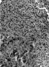
Large fragment of poorly differentiated ductal adenocarcinoma obtained using endoscopic ultrasound guided fine needle aspiration biopsy showing sheets of tumour cells with severe cytological atypia. Note the abundant fibrous stroma. Haematoxylin-eosin stain; original magnification × 360.
Figure 4
Figure 4
Cluster of monomorphous and regular small cells, without glandular or neuroendocrine structures, stained by the immunoperoxidase technique for KL1 (inset). The diagnosis of solid pseudopapillary and cystic tumour was proposed in view of the clear aspect of the cytoplasm.
Figure 5
Figure 5
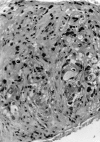
Large core biopsy illustrating a neuroendocrine tumour composed of ribbons of uniform small cells with fibrovascular stroma and staining positive for chromogranin A (inset). Haematoxylin-eosin stain; original magnification × 360; original magnification of the inset × 720.
Figure 6
Figure 6
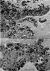
Two cases of endoscopic ultrasound guided fine needle aspiration biopsy showing small tumour fragment samples. Top, shreds of epithelial cells with moderate cytological atypia compatible with intraductal papillary mucinous tumour of the pancreas; bottom, poorly differentiated adenocarcinoma. Haematoxylin-eosin stain; original magnification × 360.

References

    1. Eur J Cancer. 1997 Apr;33(4):696-7 - PubMed
    1. Endoscopy. 1997 Jun;29(5):384-8 - PubMed
    1. Gastrointest Endosc. 1998 Feb;47(2):121-7 - PubMed
    1. Acta Cytol. 1978 Sep-Oct;22(5):327-30 - PubMed
    1. Radiology. 1979 Feb;130(2):345-6 - PubMed

Publication types

MeSH terms